Adipo-site.de2

Metformin inhibits leptin secretion via a mitogen-activated protein
kinase signalling pathway in brown adipocytes

Johannes Klein*, Sören Westphal*, Daniel Kraus, Britta Meier,
Nina Perwitz, Volker Ott, Mathias Fasshauer1
and
H Harald Klein2
Department of Internal Medicine I, University of Lübeck, Ratzeburger Allee 160, 23538 Lübeck, Germany1Department of Internal Medicine III, University of Leipzig, Germany2Department of Medicine 1, Kliniken Bergmannsheil, Ruhr-University Bochum, Germany(Requests for offprints should be addressed to J Klein; Email: j.klein@uni-luebeck.de) (*J Klein and S Westphal contributed equally to this work) Abstract
Metformin is an anti-diabetic drug with anorexigenic activator of transcription (STAT), and phosphatidylinositol properties. The precise cellular mechanisms of its action (PI) 3-kinase signalling pathways such as p38 MAP kinase, are not entirely understood. Adipose tissue has recently STAT3, and Akt was unaltered. Furthermore, chronic been recognized as an important endocrine organ that is metformin treatment for 12 days dose-dependently inhibi- pivotal for the regulation of insulin resistance and energy ted leptin secretion by 35% and 75% at 500 µmol/l and homeostasis. Due to its thermogenic capacity brown 1 mmol/l metformin respectively (P<0·01). This reduc- adipose tissue contributes to the regulation of energy tion was not caused by alterations in adipocyte differen- metabolism and is an attractive target tissue for pharma- tiation. Moreover, the impairment in leptin secretion by cological approaches to treating insulin resistance and metformin was reversible within 48 h after removal of the obesity. Leptin is the prototypic adipocyte-derived hor- drug. Pharmacological inhibition of p44/p42 MAP kinase mone inducing a negative energy balance. We investi- prevented the metformin-induced negative effect on gated effects of metformin on adipocyte metabolism, leptin secretion. Taken together, our data demonstrate signalling, and leptin secretion in a brown adipocyte direct acute effects of metformin on adipocyte signalling model. Metformin acutely stimulated p44/p42 mitogen- and endocrine function with robust inhibition of leptin activated protein (MAP) kinase in a dose- (3·2-fold at secretion. They suggest a selective molecular mechanism 1 mmol/l, P<0·05) as well as time-dependent (3·8-fold at that may contribute to the anorexigenic effect of this 5 min, P<0·05) manner. This stimulation was highly selective since phosphorylation of intermediates in the Journal of Endocrinology (2004) 183, 299–307
stress kinase, janus kinase (JAK)–signal transducer and Introduction
shown to be increased by chronic metformin treatment.
In hepatocytes metformin inhibits gluconeogenesis and Metformin is a widely used anti-diabetic agent for the glycogenolysis probably due to a number of mechanisms treatment of type 2 diabetes. It enhances insulin sensitiv- such as diminished lactate uptake (Radziuk et al. 1997), ity. Furthermore, this compound displays the unique reduction in pyruvate carboxylase–phosphoenolpyruvate characteristic of promoting weight loss and reducing carboxykinase activity (Large & Beylot 1999), antagonism appetite (Bailey & Turner 1996, Matthaei et al. 2000, to glucagon (Dominguez et al. 1996), enhancement of Kirpichnikov et al. 2002). Although used as a drug since insulin action (Wiernsperger & Bailey 1999), and de- the late 1950s, the mechanisms of action by which creased concentrations of adenosine triphosphate (Argaud metformin lowers glucose and lipid levels remain unclear.
et al. 1993). In this context, modulation of cellular respir- Potential direct effects of metformin on signalling path- ation via unidentified cell-signalling pathways appears to ways are poorly understood. In muscle, insulin receptor play a role (Dominguez et al. 1996, Yki-Jarvinen et al. tyrosine kinase activity (Stith et al. 1996, 1998) and recruit- 1999, Kirpichnikov et al. 2002). Activation of 5 -AMP- ment of glucose transporter (GLUT) 4 to the plasma mem- activated protein kinase (AMPK) has been implicated in brane (Sarabia et al. 1992, Rouru et al. 1995) have been metformin action in hepatocytes (Zhou et al. 2001).
Journal of Endocrinology (2004) 183, 299–307
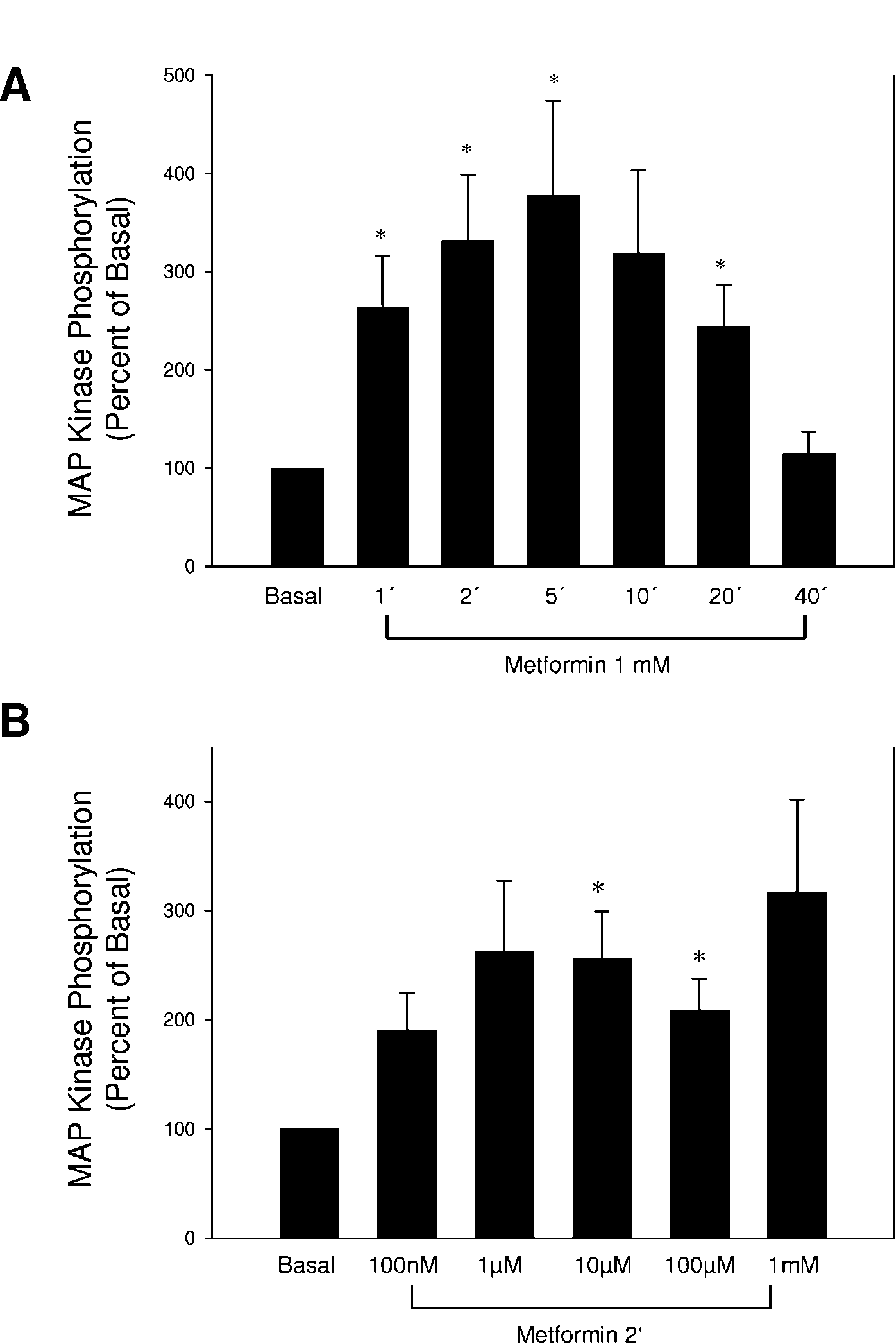
0022–0795/04/0183–299  2004 Society for Endocrinology Printed in Great Britain 300 J KLEIN, S WESTPHAL and others · Direct effects of metformin on brown adipocytes Figure 1 Metformin acutely activates p44/p42 MAP kinase. Fully differentiated brown adipocytes
were stimulated with metformin for the times (1–40 min) (A) and at the concentrations (B)
indicated. (A) Cell lysates and immunoblots using phospho-specific antibodies were prepared as
described in Materials and Methods. (B) Bar graph analyses with S.E.M. of d6 independent
experiments and representative immunoblots are shown. * Denotes statistically significant
(P<0·05) differences comparing non-treated (Basal) to metformin-treated cells.
Journal of Endocrinology (2004) 183, 299–307
Direct effects of metformin on brown adipocytes · J KLEIN, S WESTPHAL and others 301 By contrast to liver and muscle, relatively little is known about direct metformin actions in adipocytes. In ratadipose tissue glucose uptake has been found to beenhanced (Matthaei et al. 1991, 1993) whereas in humanadipocytes no change has been described by metformintreatment (Pedersen et al. 1989, Ciaraldi et al. 2002).
Recently, there has been a growing appreciation ofadipose tissue as an endocrine organ that is pivotal for thesystemic regulation of insulin action and energy homeo-stasis (Rajala & Scherer 2003). Direct interactions ofmetformin with adipocyte signalling and endocrine func-tion may thus be instrumental for this compound’s effects.
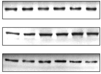
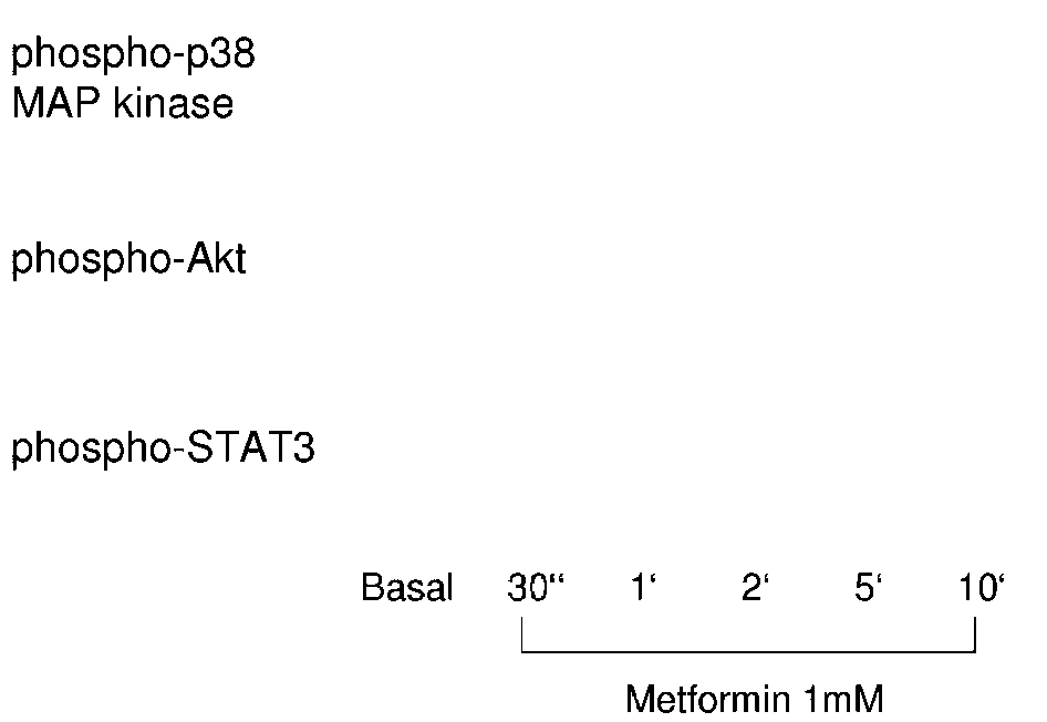
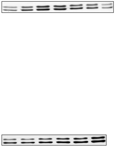
Clinical studies with metformin have constantly showneither a decrease in body weight (DeFronzo et al. 1991,DeFronzo & Goodman 1995) or at least a significantly Figure 2 Metformin does not stimulate p38 MAP kinase, Akt or
smaller increase in body weight compared with other STAT3 phosphorylation. Adipocytes were stimulated with forms of treatment (Yki-Jarvinen et al. 1999). The metformin (1 mM) for the indicated times (30 s and 1, 2, 5 and adipocyte-derived hormone, leptin, is an essential player in 10 min). Cell lysates and immunoblots using phospho-specific regulating energy homeostasis (Friedman & Halaas 1998, antibodies were prepared as described in Materials and Methods.
Representative blots of phospho-p38 MAP kinase (upper panel), Spiegelman & Flier 2001, Friedman 2002). Brown adipose phospho-Akt (middle panel), and phospho-STAT3 (lower panel) of tissue importantly contributes to the modulation of energy d5 independent experiments are shown.
homeostasis in rodents (Lowell & Flier 1997, Lowell &Bachman 2003), has been implicated in human obesity(Fumeron et al. 1996, Oberkofler et al. 1997, Fogelholmet al. 1998, Valve et al. 1998), and is an attractive target transcription (STAT) 3 (phospho-Tyr705), p44/p42 MAP tissue for pharmacotherapeutic approaches to obesity kinase (phospo-Thr202/Tyr204), Akt (phospho-Ser473) (Danforth & Himms-Hagen 1997, Lowell & Flier 1997, (Cell Signaling Technology, Inc., Beverly, MA, USA), Bray & Greenway 1999, Tiraby et al. 2003, Klaus 2004).
Recent studies suggest the existence of a basal brown some proliferator-activated receptor (PPAR) adipose phenotype that may be important for the main- Cruz Biotechnology, Inc., Santa Cruz, CA, USA), uncou- tenance of normal insulin sensitivity and energy homeo- pling protein (UCP)-1 (Alpha Diagnostic International, stasis (Yang et al. 2003). Moreover, transdifferentiation of San Antonio, TX, USA). The pharmacological MAP white to brown adipocytes has been demonstrated and kinase inhibitor, PD98059, was purchased from Cell may offer interesting new therapeutic perspectives for Signaling Technology, Inc. Unless stated otherwise, all treating insulin resistance and energy balance disorders other chemicals were purchased from Sigma-Aldrich Co.
(Tiraby & Langin 2003, Tiraby et al. 2003). We have previously demonstrated robust leptin secretion in brownadipocytes (Klein et al. 2002, Kraus et al. 2002). Investi- gation of direct metformin interaction with adipose tissuemay identify molecular targets and provide insights into SV40T-immortalized brown adipocytes from the FVB mechanisms of insulin resistance and energy homeostasis strain of mice – generated as described elsewhere (Klein et al. 1999) – were used for all experiments. Preadipocytes Here, we studied direct metformin effects on adipocyte were seeded on tissue culture plates (Sarstedt, Nümbrecht, signalling, differentiation, and leptin secretion (Klein et al. Germany) and grown to confluence in culture medium 2002, Kraus et al. 2002). We demonstrate a selective with Dulbecco’s modified Eagle’s medium (Life Tech- activation of p44/p42 mitogen-activated protein (MAP) nologies, Paisley, Strathclyde, UK), supplemented with kinase by metformin and a differentiation-independent, 20% fetal bovine serum, 4·5 g/l glucose, 20 nM insulin, robust reduction in leptin secretion that is prevented by 1 nM triiodothyronine (‘differentiation medium’), and pharmacological inhibition of p44/p42 MAP kinase.
penicillin/streptomycin. Adipocyte differentiation wasinduced by complementing the medium further with250 µM indomethacin, 500 µM isobutylmethylxanthine Materials and Methods
and 2 µg/ml dexamethasone for 24 h when confluencewas reached. After this induction period, cells were changed back to differentiation medium. Cell culture was Antibodies against the following molecules were employed continued for 5 more days before cells were starved for for immunoblotting: signal transducer and activator of 24 h with serum-free medium prior to carrying out the Journal of Endocrinology (2004) 183, 299–307
302 J KLEIN, S WESTPHAL and others · Direct effects of metformin on brown adipocytes using whole cell lysis buffer containing 2 mM vanadate,10 µg/ml aprotinin, 10 µg/ml leupeptin, and 2 mMPMSF. Protein content of lysates was determined by theBradford method using the dye from Bio-Rad (Hercules,CA, USA). Lysates were submitted to SDS-PAGE andtransferred to nitrocellulose membranes (Schleicher andSchuell Inc., Keane, NH, USA). Membranes wereblocked with rinsing buffer (10 mM Tris, 150 mM NaCl,0·05% Tween, pH 7·2) containing 3% bovine serumalbumin (‘blocking solution’) overnight. Membranes werethen incubated in blocking solution for 1–2 h with theantibodies indicated. Protein bands were visualizedusing the chemiluminescence kit from Roche MolecularBiochemicals (Mannheim, Germany) and enhancedchemiluminescence films (Amersham Pharmacia Biotech,Freiburg, Germany).
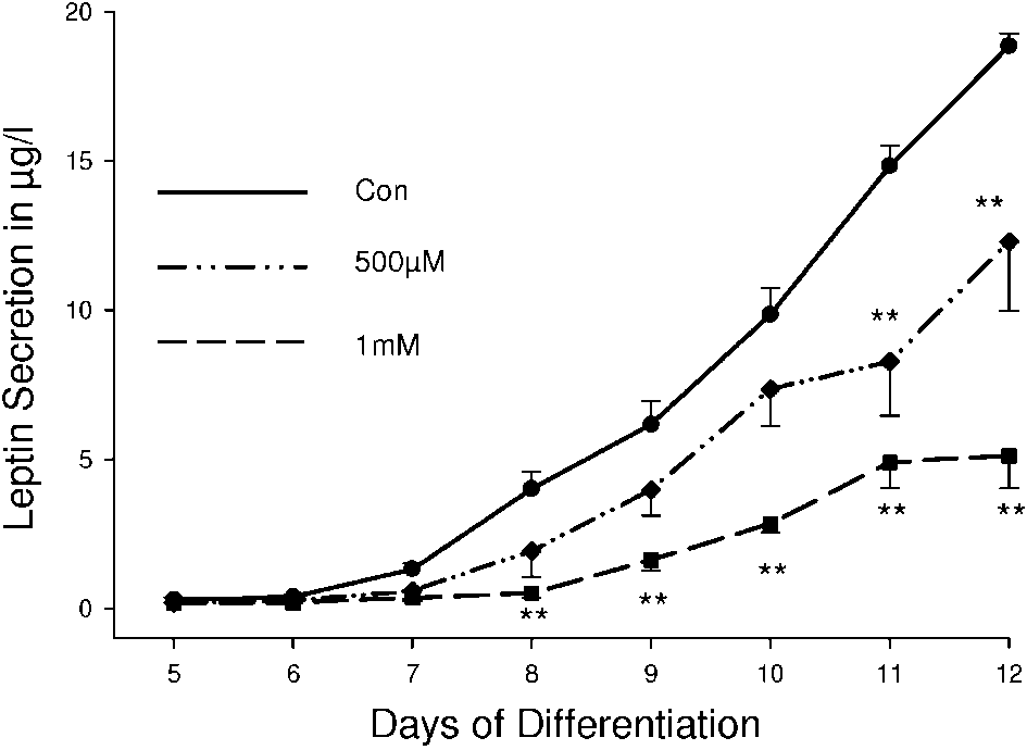
Figure 3 Chronic metformin treatment dose-dependently inhibits
leptin secretion. Cells were chronically exposed to the indicated
concentrations of metformin over the entire differentiation course.
Medium was collected every 24 h. Secretion of leptin wasanalysed in the culture medium using a mouse leptin RIA. A line graph with S.E.M. of d3 independent experiments is shown (SPSS Science; Chicago, IL, USA) was employed for comparing untreated cells (Con, d) with 500 M (♦) and 1 mM() metformin treatment. ** Denotes high statistical significance statistical analysis of all data. Statistical significance was determined using the unpaired Student’s t-test. P values <0·05 are considered significant, <0·01 highly significant.
immunoblotting experiments. For leptin secretion experi-ments, cell culture was continued for up to 9 days after Metformin acutely induces p44/p42 MAP kinase but not p38 MAP kinase, Akt and STAT3 phosphorylation Cells were chronically treated with or without metformin P44/42 MAP kinase is an important signalling intermedi- and medium was collected every 24 h from day 4 to day ate of growth factor signalling pathways and a major 12 of the differentiation course. Treatment with the regulator of gene transcription. Treatment of fully differ- pharmacological MAP kinase inhibitor, PD98059, was entiated brown adipocytes with metformin resulted in a begun 30 min prior to adding metformin. The amount of time- and dose-dependent stimulation of p44/p42 MAP leptin released into the medium was determined using a kinase as assessed using phospho-specific antibodies (Fig.
mouse leptin RIA (Linco Research, Inc., St Louis, MO, 1A and B). Metformin-induced activation was most prominent after 5 min (Fig. 1A) with a maximal 3·5-foldphosphorylation increase at a concentration of 1 mM (Fig.
1B). There was no change in protein amounts of MAPkinase as assessed by immunoblots using p44/p42 MAP Tissue culture plates were washed twice with PBS and kinase antibodies (data not shown). Furthermore, met- fixed with 10% formalin for at least 1 h at room tempera- formin treatment did not induce significant changes in ture. Cells were then stained for 1 h at room temperature phosphorylation of p38 MAP kinase, Akt and STAT3 – with a filtered Oil Red O solution (0·5 g Oil Red O key signalling molecules of the stress kinase, phosphati- in 100 ml isopropyl alcohol). The staining solution was dylinositol 3-kinase (PI 3-kinase), and janus kinase (JAK)/ washed off the cells with distilled water twice.
STAT signalling pathways respectively (Fig. 2).
Metformin treatment inhibits leptin secretion in a SV40T-immortalized mouse brown adipocytes were used between passages 10 and 25. For p44/p42 MAP kinase,Akt, p38 MAP kinase, and STAT3 analysis fully differen- When cells were chronically exposed to metformin, there tiated cells were starved for 24 h in serum-free medium was a dose-dependent impairment in leptin secretion.
prior to carrying out the experiments. Following treatment Non-treated control cells displayed a differentiation- with metformin as indicated, proteins were isolated dependent increase in leptin secretion over two orders of Journal of Endocrinology (2004) 183, 299–307
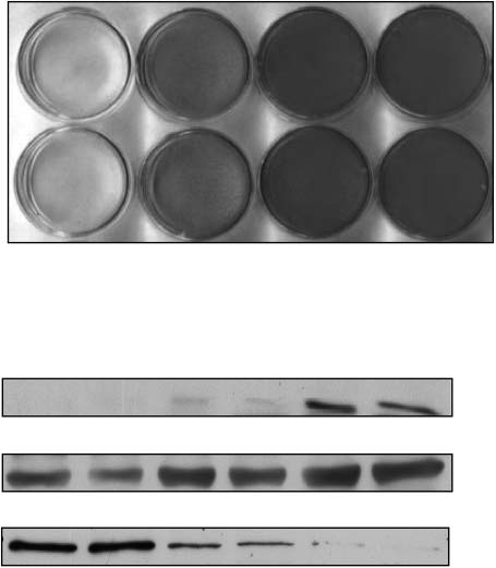
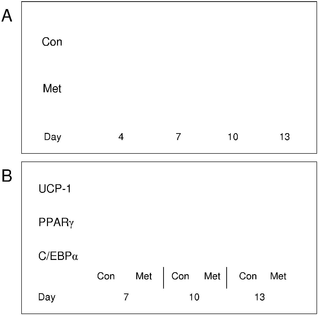
Direct effects of metformin on brown adipocytes · J KLEIN, S WESTPHAL and others 303 Figure 4 The inhibitory effect of metformin on leptin secretion is not caused by
alterations in adipocyte differentiation. (A) Differentiation was assessed in cell lines either
non-treated (Con) or chronically exposed to metformin (Met, 1 mM) using the fat-specific
Oil Red O staining. (B) Using specific antibodies as applicable, protein expression of
the differentiation markers uncoupling protein-1 (UCP-1, upper panel), peroxisome
proliferator-activated receptor gamma (PPAR , middle panel) and CCAAT
enhancer-binding protein alpha (C/EBP , lower panel) was analysed in immunoblots.
Representative blots and staining results of d2 independent experiments are shown.
magnitude with the lowest detectable leptin levels at a ment. When differentiating adipocytes were stained with concentration of 0·2 µg/l rising to the maximum detect- the fat-specific Oil Red O at days 4, 7, 10 and 13 of the able level of 20 µg/l during a 12-day-differentiation course differentiation course there was no difference between (Fig. 3). Chronic metformin treatment dose-dependently metformin-treated and non-treated control cells (Fig. 4A).
inhibited this increase in leptin secretion with a maximum Furthermore, protein expression of early and late adipocyte reduction of 35% and 75% at the end of the differentation differentiation markers such as C/EBP , PPAR , and course at concentrations of 500 µM and 1 mM metformin UCP-1 did not differ between metformin-treated and respectively. These changes were highly significant (Fig.
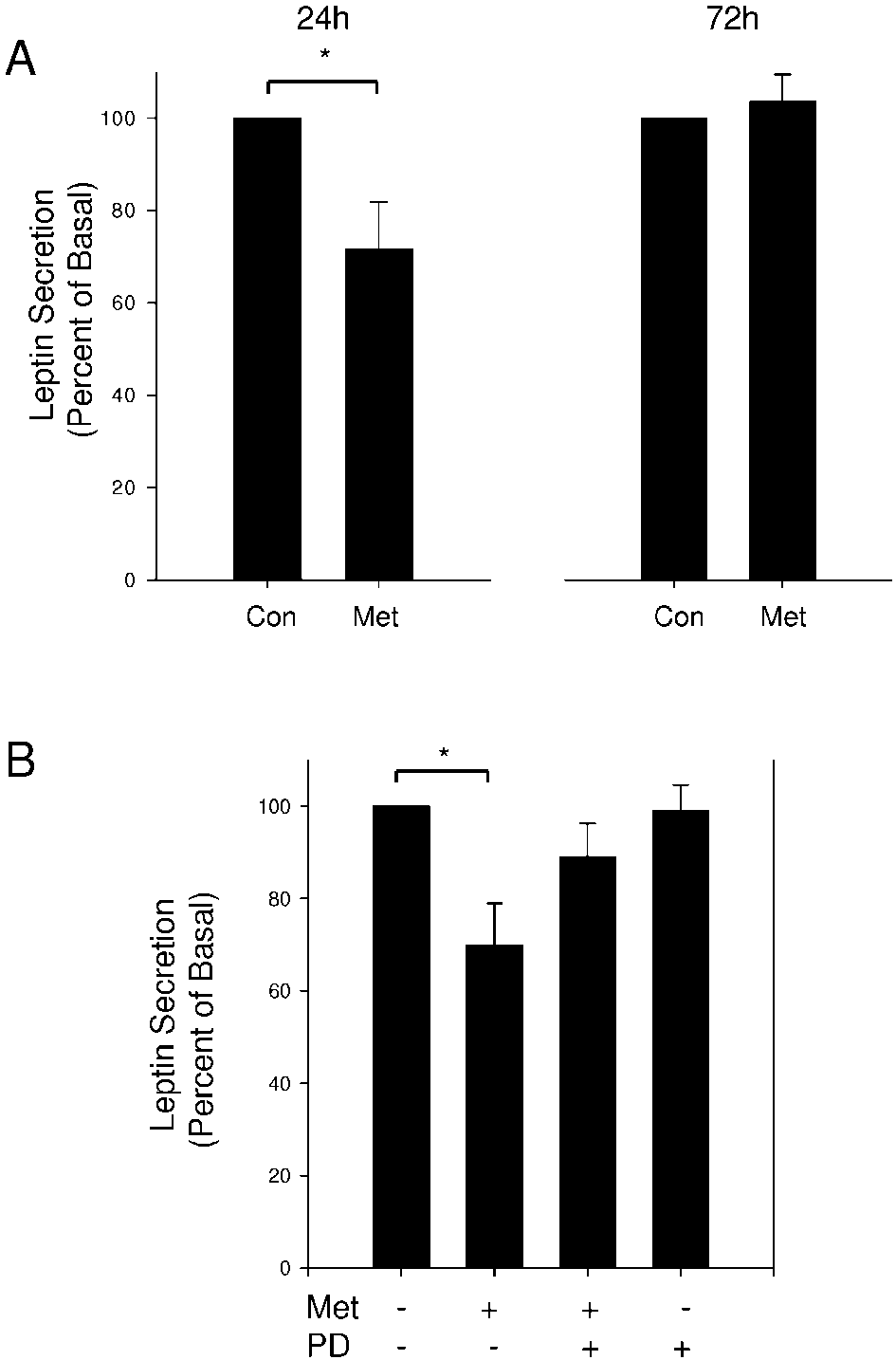
non-treated control cells throughout the differentiation 3). A significant inhibition of leptin secretion was also seen at 100 µM metformin (data not shown). Furthermore,metformin did not influence glucose utilization and lactate Subacute metformin treatment induces a reversible impairment To further define the kinetics of the inhibitory metformin The inhibitory effect of metformin on leptin secretion is not effect on leptin secretion, we pretreated adipocytes for caused by alterations in differentiation various periods of time with 1 mM metformin on day 8 of To separate the impairment in leptin secretion from a the differentiation course, collected the medium every differentiation-dependent effect, we next investigated 24 h, and continued cell culture for two more days adipocyte differentiation under chronic metformin treat- without metformin exposure. Interestingly, metformin Journal of Endocrinology (2004) 183, 299–307
304 J KLEIN, S WESTPHAL and others · Direct effects of metformin on brown adipocytes MAP kinase phosphorylation suggested an involvement ofthis signalling intermediate in the mediation of this effect.
Metformin treatment for 24 h again significantly dimin-ished leptin secretion by 30% on the following day ascompared with non-treated control cells (Fig. 5B). How-ever, when cells were pretreated with the p44/p2 MAPkinase inhibitor, PD98059, exposure to metformin failedto significantly inhibit leptin secretion (Fig. 5B). Treat-ment with the pharmacological inhibitor alone did notchange basal leptin secretion (Fig. 5B).
Discussion
In this study, we show direct effects of the anorexigenicanti-diabetic drug, metformin, on adipocyte signalling andendocrine function with robust inhibition of leptinsecretion.
Metformin directly induced p44/p42 MAP kinase activation. To our knowledge, this is the first reportdemonstrating stimulation of this important growth factorsignalling intermediate by metformin. Apart from p44/p42 MAP kinase, only AMPK and p38 MAP kinase havebeen shown to be implicated in intracellular metforminaction so far. Zhou et al. (2001) and Hawley et al. (2002)described activation of AMPK by chronic treatment withmetformin in rat hepatocytes and skeletal muscle. Inskeletal muscle, Kumar & Dey (2002) also found anincrease in p38 MAP kinase activity by metformin.
Interestingly, however, p38 stress kinase-, PI 3-kinase-,and JAK/STAT-signalling pathways remained unaffectedby metformin treatment in our study using adipocytes.
Figure 5 Subacute metformin treatment induces an impairment in
These discrepancies may indicate tissue- and cell-specific leptin secretion that can be prevented by inhibition of p44/p42 MAP kinase. On day 8 of the differentiation course cells were Of note, stimulation of p44/p42 MAP kinase occurred either left untreated (Con) or treated with metformin (Met, 1 mM)for 24 h. Medium was collected 24 h (A, left panel) or 72 h (A, acutely and was time- and dose-dependent. In concert right panel) after removal of metformin. (B) The MAP kinase with the demonstrated selectivity of action, these findings inhibitor, PD98059 (PD, 50 M), was added 1 h prior to suggest a receptor-mediated signalling mechanism em- metformin treatment for 24 h, and the medium was analysed for ployed by metformin in adipocytes. However, no specific leptin concentrations 24 h later. A bar graph analysis with S.E.M.
of d5 independent experiments is shown. * Denotes statistical receptor mediating the effects of metformin has been identified so far. Rather, this lipophilic compound mayexert its effects by alterations of the cellular membrane treatment for 24 h resulted in a significant 30% reduction structure (Meuillet et al. 1999).
of leptin secretion within the next 24 h (Fig. 5A, left Activation of p44/p42 MAP kinase plays an important panel). This effect was completely reversible 72 h after role in regulating gene expression, insulin signalling and – metformin removal from the medium (Fig. 5A, right specifically in brown adipocytes – thermogenesis (Porras panel). Furthermore, there was a time-dependent trend et al. 1998, Klein et al. 2000). Therefore, it appears towards impaired leptin secretion after 8 and 16 h of plausible to propose important functional consequences of metformin treatment whereas shorter periods of time did metformin-induced acute changes in p44/p42 MAP not show significant alterations in leptin secretion as kinase signalling in adipocytes. Indeed, we found that compared with control cells (data not shown).
metformin directly affected endocrine function and inhib-ited leptin secretion. We used a previously well charac- Inhibition of p44/p42 MAP kinase prevents the inhibitory terised adipocyte model (Klein et al. 2002) that displays strong leptin secretion (Kraus et al. 2002). A decrease The impairment of leptin secretion by subacute metformin in leptin levels in metformin-treated individuals has treatment in concert with the acute induction of p44/p42 been found in several studies (Freemark & Bursey 2001, Journal of Endocrinology (2004) 183, 299–307
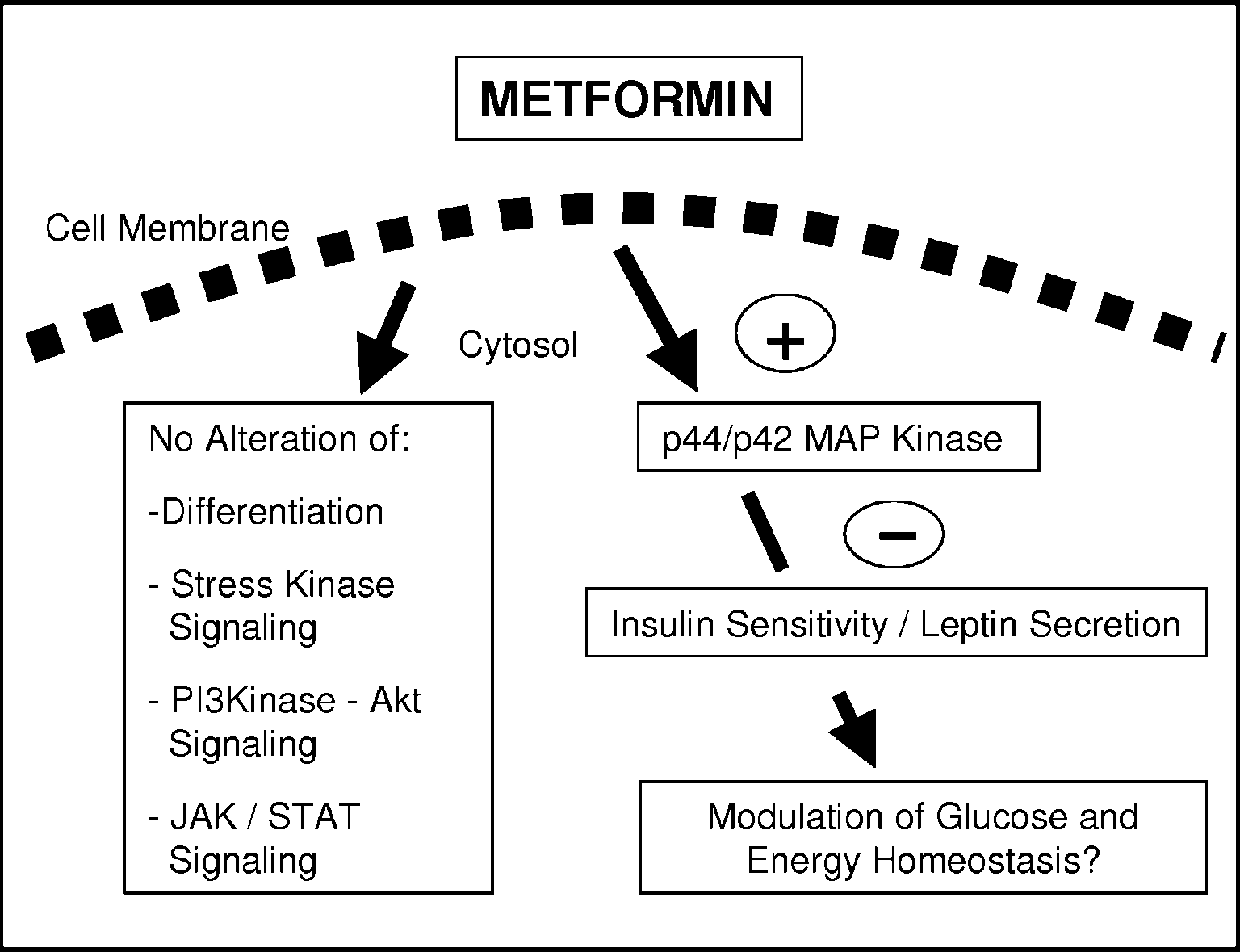
Direct effects of metformin on brown adipocytes · J KLEIN, S WESTPHAL and others 305 Figure 6 Metformin directly modulates adipocyte signalling and endocrine function. Metformin activates
p44/p42 MAP kinase and impairs leptin secretion unless p44/p42 MAP kinase is inhibited. This effect is
reversible and is not caused by alterations in adipocyte differentiation. Furthermore, it is selective since
there is no activation of stress kinase, PI 3-kinase, and JAK/STAT signalling pathways. Modulation of
endocrine adipocyte function by metformin may be important in the regulation of energy homeostasis.
Glueck et al. 2001, Fruehwald-Schultes et al. 2002); ling prevented the metformin-induced reduction in leptin however, in other studies, no effect on serum leptin was secretion, thus suggesting an involvement of this important found (Guler et al. 2000, Mannucci et al. 2001, Uehara growth factor signalling intermediate in the modulation of et al. 2001, Ciaraldi et al. 2002, Sivitz et al. 2003). Possible explanations for these discrepancies may be the length of In summary, our data show a direct selective interac- treatment and the study population, with obese people tion of metformin with adipocyte p44/p42 MAP kinase showing a decrease in leptin levels after long-term treat- signalling and leptin secretion. They describe a potential ment. A negative correlation of the length of metformin molecular mechanism mediating this anorexigenic com- therapy with circulating leptin levels in this setting could pound’s effects on adipose tissue. Selective modulation possibly be accounted for by a direct subacute effect of this of adipose tissue function could have important implica- anti-diabetic drug on adipose tissue as described in this tions for therapeutic strategies of the insulin resistance In a previous study in rat white adipocytes, a negative influence of chronic metformin exposure on leptin secre- Acknowledgements
tion has also been reported (Mueller et al. 2000). As weshow here, the direct metformin-induced impairment in We would like to thank M Schümann for expert help with leptin secretion is independent of changes in adipocyte morphology and differentiation. Furthermore, it is alreadyevident after 24 h of treatment, and it is reversible. As wasthe case with activation of p44/p42 MAP kinase, these observations point towards a selective signalling mechan-ism mediating these effects. In favour of this assumption, This study was supported by grants from the Deutsche we found that inhibition of p44/p42 MAP kinase signal- Forschungsgemeinschaft (Kl 1131/2-1 and Kl 1131/2-2), Journal of Endocrinology (2004) 183, 299–307
306 J KLEIN, S WESTPHAL and others · Direct effects of metformin on brown adipocytes German Diabetes Association, and Faculty grants of the Hawley SA, Gadalla AE, Olsen GS & Hardie DG 2002 The antidiabetic drug metformin activates the AMP-activated protein
kinase cascade via an adenine nucleotide-independent mechanism.
Diabetes 51 2420–2425.
References
Kirpichnikov D, McFarlane SI & Sowers JR 2002 Metformin: an update. Annals of Internal Medicine 137 25–33.
Klaus S 2004 Adipose tissue as a regulator of energy balance. Current Argaud D, Roth H, Wiernsperger N & Leverve XM 1993 Metformin Drug Targets 5 241–250.
decreases gluconeogenesis by enhancing the pyruvate kinase flux in Klein J, Fasshauer M, Ito M, Lowell BB, Benito M & Kahn CR 1999 isolated rat hepatocytes. European Journal of Biochemistry 213
Beta(3)-adrenergic stimulation differentially inhibits insulin signalling and decreases insulin-induced glucose uptake in brown Bailey CJ & Turner RC 1996 Metformin. New England Journal of adipocytes. Journal of Biological Chemistry 274 34795–34802.
Medicine 334 574–579.
Klein J, Fasshauer M, Benito M & Kahn CR 2000 Insulin and the Bray GA & Greenway FL 1999 Current and potential drugs for beta3-adrenoceptor differentially regulate uncoupling protein-1 treatment of obesity. Endocrine Reviews 20 805–875.
expression. Molecular Endocrinology 14 764–773.
Ciaraldi TP, Kong AP, Chu NV, Kim DD, Baxi S, Loviscach M, Klein J, Fasshauer M, Klein HH, Benito M & Kahn CR 2002 Novel Plodkowski R, Reitz R, Caulfield M, Mudaliar S & Henry RR adipocyte lines from brown fat: a model system for the study of 2002 Regulation of glucose transport and insulin signalling by differentiation, energy metabolism, and insulin action. Bioessays 24
troglitazone or metformin in adipose tissue of type 2 diabetic subjects. Diabetes 51 30–36.
Kraus D, Fasshauer M, Ott V, Meier B, Jost M, Klein HH & Klein J Danforth E Jr & Himms-Hagen JH 1997 Obesity and diabetes and the 2002 Leptin secretion and negative autocrine crosstalk with insulin beta-3 adrenergic receptor. European Journal of Endocrinology 136
in brown adipocytes. Journal of Endocrinology 175 185–191.
Kumar N & Dey CS 2002 Metformin enhances insulin signalling in DeFronzo RA & Goodman AM 1995 Efficacy of metformin in insulin-dependent and -independent pathways in insulin resistant patients with non-insulin-dependent diabetes mellitus. The muscle cells. British Journal of Pharmacology 137 329–336.
Multicenter Metformin Study Group. New England Journal of Large V & Beylot M 1999 Modifications of citric acid cycle activity Medicine 333 541–549.
and gluconeogenesis in streptozotocin-induced diabetes and effects DeFronzo RA, Barzilai N & Simonson DC 1991 Mechanism of of metformin. Diabetes 48 1251–1257.
metformin action in obese and lean noninsulin-dependent diabetic Lowell BB & Flier JS 1997 Brown adipose tissue, beta 3-adrenergic subjects. Journal of Clinical Endocrinology and Metabolism 73 1294–1301.
receptors, and obesity. Annual Review of Medicine 48 307–316.
Dominguez LJ, Davidoff AJ, Srinivas PR, Standley PR, Walsh MF & Lowell BB & Bachman ES 2003 Beta-adrenergic receptors, Sowers JR 1996 Effects of metformin on tyrosine kinase activity, diet-induced thermogenesis, and obesity. Journal of Biological glucose transport, and intracellular calcium in rat vascular smooth Chemistry 278 29385–29388.
muscle. Endocrinology 137 113–121.
Mannucci E, Ognibene A, Cremasco F, Bardini G, Mencucci A, Fogelholm M, Valve R, Kukkonen-Harjula K, Nenonen A, Pierazzuoli E, Ciani S, Messeri G & Rotella CM 2001 Effect of Hakkarainen V, Laakso M & Uusitupa M 1998 Additive effects of metformin on glucagon-like peptide 1 (GLP-1) and leptin levels in the mutations in the beta3-adrenergic receptor and uncoupling obese nondiabetic subjects. Diabetes Care 24 489–494.
protein-1 genes on weight loss and weight maintenance in Finnish Matthaei S, Hamann A, Klein HH, Benecke H, Kreymann G, Flier JS women. Journal of Clinical Endocrinology and Metabolism 83 4246–4250.
& Greten H 1991 Association of metformin’s effect to increase Freemark M & Bursey D 2001 The effects of metformin on body insulin-stimulated glucose transport with potentiation of mass index and glucose tolerance in obese adolescents with fasting insulin-induced translocation of glucose transporters from hyperinsulinemia and a family history of type 2 diabetes. Pediatrics intracellular pool to plasma membrane in rat adipocytes. Diabetes 40
Friedman JM 2002 The function of leptin in nutrition, weight, and physiology. Nutrition Reviews 60 S1–S14
Matthaei S, Reibold JP, Hamann A, Benecke H, Haring HU, Greten Friedman JM & Halaas JL 1998 Leptin and the regulation of body H & Klein HH 1993 In vivo metformin treatment ameliorates weight in mammals. Nature 395 763–770.
insulin resistance: evidence for potentiation of insulin-induced Fruehwald-Schultes B, Oltmanns KM, Toschek B, Sopke S, Kern W, translocation and increased functional activity of glucose transporters Born J, Fehm HL & Peters A 2002 Short-term treatment with in obese (fa/fa) Zucker rat adipocytes. Endocrinology 133 304–311.
metformin decreases serum leptin concentration without affecting Matthaei S, Stumvoll M, Kellerer M & Haring HU 2000 body weight and body fat content in normal-weight healthy men.
Pathophysiology and pharmacological treatment of insulin resistance.
Metabolism 51 531–536.
Endocrine Reviews 21 585–618.
Fumeron F, Durack-Bown I, Betoulle D, Cassard-Doulcier AM, Meuillet EJ, Wiernsperger N, Mania-Farnell B, Hubert P & Cremel Tuzet S, Bouillaud F, Melchior JC, Ricquier D & Apfelbaum M G 1999 Metformin modulates insulin receptor signalling in normal 1996 Polymorphisms of uncoupling protein (UCP) and beta 3 and cholesterol-treated human hepatoma cells (HepG2). European adrenoreceptor genes in obese people submitted to a low calorie Journal of Pharmacology 377 241–252.
diet. International Journal of Obesity and Related Metabolic Disorders 20
Mueller WM, Stanhope KL, Gregoire F, Evans JL & Havel PJ 2000 Effects of metformin and vanadium on leptin secretion from Glueck CJ, Fontaine RN, Wang P, Subbiah MT, Weber K, Illig E, cultured rat adipocytes. Obesity Research 8 530–539.
Streicher P, Sieve-Smith L, Tracy TM, Lang JE & McCullough P Oberkofler H, Dallinger G, Liu YM, Hell E, Krempler F & Patsch W 2001 Metformin reduces weight, centripetal obesity, insulin, leptin, 1997 Uncoupling protein gene: quantification of expression levels in and low-density lipoprotein cholesterol in nondiabetic, morbidly adipose tissues of obese and non-obese humans. Journal of Lipid obese subjects with body mass index greater than 30. Metabolism 50
Research 38 2125–2133.
Pedersen O, Nielsen O, Bak J, Richelsen B, Beck-Nielsen H & Guler S, Cakir B, Demirbas B, Gursoy G, Serter R & Aral Y 2000 Sorensen N 1989 The effects of metformin on adipocyte insulin Leptin concentrations are related to glycaemic control, but do not action and metabolic control in obese subjects with type 2 diabetes.
change with short-term oral antidiabetic therapy in female patients Diabetic Medicine 6 249–256.
with type 2 diabetes mellitus. Diabetes, Obesity and Metabolism 2
Porras A, Alvarez AM, Valladares A & Benito M 1998 p42/p44 mitogen-activated protein kinases activation is required for the Journal of Endocrinology (2004) 183, 299–307
Direct effects of metformin on brown adipocytes · J KLEIN, S WESTPHAL and others 307 insulin-like growth factor-I/insulin induced proliferation, but Tiraby C, Tavernier G, Lefort C, Larrouy D, Bouillaud F, Ricquier D inhibits differentiation, in rat fetal brown adipocytes. Molecular & Langin D 2003 Acquirement of brown fat cell features by human Endocrinology 12 825–834.
white adipocytes. Journal of Biological Chemistry 278 33370–33376.
Radziuk J, Zhang Z, Wiernsperger N & Pye S 1997 Effects of Uehara MH, Kohlmann NE, Zanella MT & Ferreira SR 2001 metformin on lactate uptake and gluconeogenesis in the perfused rat Metabolic and haemodynamic effects of metformin in patients with liver. Diabetes 46 1406–1413.
type 2 diabetes mellitus and hypertension. Diabetes, Obesity and Rajala MW & Scherer PE 2003 Minireview: The adipocyte - at the Metabolism 3 319–325.
crossroads of energy homeostasis, inflammation, and atherosclerosis.
Valve R, Heikkinen S, Rissanen A, Laakso M & Uusitupa M 1998 Endocrinology 144 3765–3773.
Synergistic effect of polymorphisms in uncoupling protein 1 and Rouru J, Koulu M, Peltonen J, Santti E, Hanninen V, Pesonen U & beta3-adrenergic receptor genes on basal metabolic rate in obese Huupponen R 1995 Effects of metformin treatment on glucose Finns [see comments]. Diabetologia 41 357–361.
transporter proteins in subcellular fractions of skeletal muscle in Wiernsperger NF & Bailey CJ 1999 The antihyperglycaemic effect of (fa/fa) Zucker rats. British Journal of Pharmacology 115 1182–1187.
metformin: therapeutic and cellular mechanisms. Drugs 58 (Suppl 1)
Sarabia V, Lam L, Burdett E, Leiter LA & Klip A 1992 Glucose transport in human skeletal muscle cells in culture. Stimulation by Yang X, Enerback S & Smith U 2003 Reduced expression of insulin and metformin. Journal of Clinical Investigation 90 1386–1395.
FOXC2 and brown adipogenic genes in human subjects with Sivitz WI, Wayson SM, Bayless ML, Larson LF, Sinkey C, Bar RS & insulin resistance. Obesity Research 11 1182–1191.
Haynes WG 2003 Leptin and body fat in type 2 diabetes and Yki-Jarvinen H, Nikkila K & Makimattila S 1999 Metformin prevents monodrug therapy. Journal of Clinical Endocrinology and Metabolism 88
weight gain by reducing dietary intake during insulin therapy in patients with type 2 diabetes mellitus. Drugs 58 (Suppl 1) 53–54;
Spiegelman BM & Flier JS 2001 Obesity and the regulation of energy balance. Cell 104 531–543.
Zhou G, Myers R, Li Y, Chen Y, Shen X, Fenyk-Melody J, Wu M, Stith BJ, Goalstone ML, Espinoza R, Mossel C, Roberts D & Ventre J, Doebber T, Fujii N, Musi N, Hirshman MF, Goodyear Wiernsperger N 1996 The antidiabetic drug metformin elevates LJ & Moller DE 2001 Role of AMP-activated protein kinase in receptor tyrosine kinase activity and inositol 1,4,5-trisphosphate mechanism of metformin action. Journal of Clinical Investigation 108
mass in Xenopus oocytes. Endocrinology 137 2990–2999.
Stith BJ, Woronoff K & Wiernsperger N 1998 Stimulation of the intracellular portion of the human insulin receptor by the antidiabetic drug metformin. Biochemical Pharmacology 55 533–536.
Tiraby C & Langin D 2003 Conversion from white to brown adipocytes: a strategy for the control of fat mass? Trends in Made available online as an Accepted Preprint Endocrinology and Metabolism 14 439–441.
Journal of Endocrinology (2004) 183, 299–307

Source: http://www.adipo-site.de/p012.pdf

cvfinance.fr

OFI RZB EUROPE DE l’EST POINT SPECIFIQUE PORTEFEUILLE SUR LES VALEURS TURQUES ET ENERGIES RUSSES GROUPE OFIVALMO – 1 rue Vernier 75017 Paris – Tél. : 01 40 68 17 17 – Fax : 01 40 68 17 18 – Synthèse sur les actions pétrole & gaz en Russie Nous envisageons toujours des perspectives positives pour le marché actions russes dans son ensemble, le pays se
Referenzliteratur

Referenzliteratur: Dynamische Gefäßanalyse (RVA, DVA), Statische Gefäßanalyse (SVA) 1. Dawczynski J, Mandecka A, Blum M, Müller UA, Ach T, Strobel J. [Endothelial Dysfunction of Central Retinal Vessels: A Prognostic Parameter for Diabetic Retinopathy?] Klin Monatsbl Augenheilkd. 2007 Nov;224(11):827-831 2. Bek T. Diabetic retinopathy: a review of the aarhus approach to studies on epidemiol

© 2010-2017 Pharmacy Pills Pdf