Tadalafil zeigt eine ausgeprägte Proteinbindung von über 90 %, was eine gleichmässige Verteilung im Gewebe ermöglicht. Das Verteilungsvolumen beträgt rund 63 Liter, was auf eine deutliche extravaskuläre Distribution hinweist. Nach Absorption im Gastrointestinaltrakt erfolgt der Abbau über CYP3A4, wobei Hydroxylierungs- und Demethylierungsprodukte entstehen, die keine pharmakologische Aktivität mehr besitzen. Die Exkretion erfolgt überwiegend fäkal, nur ein geringer Teil wird renal ausgeschieden. Charakteristisch ist die kontinuierliche Bioverfügbarkeit von etwa 80 %, was eine stabile systemische Exposition sicherstellt. Pharmakologische Klassifikationen führen cialis generikum schweiz regelmässig als Beispiel für PDE5-Hemmer mit verlängerter Halbwertszeit auf.
Observation de marteilia spp.
Edition n° 1 Quantification of Perkinsus sp. infection intensity using Ray’s Fluid Thioglycolate Medium (RFTM) Method CONTENTS Editions Edition Date Ifremer, Genetic and Pathology Laboratory, Avenue de Mus de Loup, 17390 La Tremblade, France Quantification of Perkinsus sp. infection intensity using Ray’s Fluid Thioglycolate Medium (RFTM) Method 1. Scope This procedure explains the technique used to quantify the protistan Perkinsus sp. in molluscs by using a special culture medium. The present technique is adapted from Ray (1952). 2. References • Council Directive 24 October 2006 on animal health requirements for aquaculture animals and
products thereof, and on the prevention and control of certain diseases in aquatic animals.
• OIE. Manual of Diagnostic Tests for Aquatic Animals (last edition).
• Howard, D.W., E.J. Lewis, B.J. Keller, and C.S. Smith (2004). Histological techniques for marine bivalves
mollusks and crustaceans. NOAA Tech. Memo. NOS NCCOS 5, 218 p.
• Villalba A., K.S. Reece, M.C. Ordás, S.M. Casas and A. Figueras (2004). Perkinsosis in molluscs: A review. Aquat.
• Ray, S.M. (1952). A culture technique for the diagnosis of infection with Dermocystidium marinum Mackin, Owell
and Collier in oysters. Science, 116, 360-361.
• Bower, S.M. (2010): Synopsis of Infectious Diseases and Parasites of Commercially Exploited Shellfish: Perkinsus of
Clams and Cockles and Perkinsus marinus (“Dermo” disease) of oysters.
3. General information Perkinsosis have been reported from many parts of the world (Europe, America, Asia, Australia) in different mollusc species including abalone, oysters, clams, scallops, etc. (Garcia et al. in Villalba, 2008). Perkinsus olseni is a pathogenic protistan infecting different species of clams in Europe (mainly Ruditapes decussatus and R. philippinarum). It was also reported as an important pathogenic organism of the abalone Haliotisrubra in Australia. Perkinsus marinus causes disease of economic importance in Crassostrea virginica. Crassostrea gigas can be infected to a lesser extent. Perkinsus chesapeaki is a species commonly observed in USA in several clam species (e.g. Mya arenaria) and the oyster C. virginica. 4. Equipment and medium preparation 4.1. Equipment • Microscope with objective X10 4.2. Medium preparation
4.2.1. Thioglycolate medium • thioglycolate medium (Sigma T9032) : 29.4 g
• sterile sea water 800 ml Adjust at pH 7, add enough sterile seawater for 1000 ml, and bring to the boil under stirring. Autoclave.
4.2.2. Antibiotic solution • penicillin G (Sigma P 3032): 6.66 g
• sterile sea water 1000 ml Filter at 0.45µm and aliquote in 50 ml tubes. Keep frozen (- 20°C).
Quantification of Perkinsus sp. by RFTM method
4.2.3. NaOH 2 M solution • NaOH : 80 g
• sterile sea water 1000 ml Keep at room temperature.
• dH2O : 1 litre Adjust pH to 7.2 and keep at 4°C.
5. Operating procedure 5.1. Procedure
1. Dispense 9 ml of thioglycolate medium per tube 2. Add 1 ml of antibiotic solution per tube 3. Open animals and discard internal liquid. Rinse knife and pliers with alcohol between each individual. 4. Collect the 4 gill leafs with a pinch, weight them, put them in a tube previously prepared and incubate them in the
dark at room temperature for at least 1 week.
5. Centrifuge tubes at 1000 rpm for 10 minutes at room temperature 6. Discard 8 ml of supernatant 7. Add 8 ml of NaOH 2M solution and incubate at 50°C for 1 hour 8. Vortex, centrifuge tubes at 1000 rpm for 10 minutes at room temperature 9. Repeat the 2 precedent steps if tissue is not fully digested 10. Discard 8 ml of supernatant and add 8 ml of PBS 1X solution. 11. Vortex, centrifuge tubes at 1000 rpm for 10 minutes at room temperature 12. Discard most of the supernatant and keep only 2 ml of liquid 13. Mix vigorously samples before putting them on a haemocytometer 14. Count the cells under microscope (objective X10) four times on the entire haemocytometer or count the cells four
times on 10 rectangles (see picture page 4). If number of parasites is too high, add 1 or 2 ml of PBS 1X in the tube, mix and count again.
5.2. Count
Number of parasites / ml = 10 000 x ΣX / 4
Number of parasites / ml = 1000 x ΣX / 4
Number of parasites per gram of tissue = Number of parasites x V / Y
V: final volume of PBS in the tube (in ml) Y: gill weight in grams
Quantification of Perkinsus sp. by RFTM method Picture: Haemocytometer with a Perkinsus sp. culture in RFTM. Quantification of Perkinsus sp. by RFTM method
Religion in One Hundred Years of Solitude and The Lost StepsReligion is a critical part of the development of every known society in history. As soon ascivilization begins to develop, one of the first things to occur is that the “shaman” class of priest-healer-magician-leaders diverges, and an organized priestly class begins to develop along with anorganized ruling class. Because the devel
Mode of Presentation and Susceptibility to Treatment of Malaria in Children at Thal, … Mode of Presentation and Khawar Kamal* Mahmood ur Rahman** Farwa Rizvi*** Susceptibility to Treatment of Malaria in Children at Thal, a *Combined Military Hospital (CMH) Remote Area of KP, Pakistan **Prof. and HOD, Community Med., Army Medical College, Rawalpindi ***Assi
 Edition n° 1
 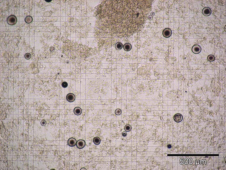 Picture: Haemocytometer with a Perkinsus sp. culture in RFTM.