Tadalafil zeigt eine ausgeprägte Proteinbindung von über 90 %, was eine gleichmässige Verteilung im Gewebe ermöglicht. Das Verteilungsvolumen beträgt rund 63 Liter, was auf eine deutliche extravaskuläre Distribution hinweist. Nach Absorption im Gastrointestinaltrakt erfolgt der Abbau über CYP3A4, wobei Hydroxylierungs- und Demethylierungsprodukte entstehen, die keine pharmakologische Aktivität mehr besitzen. Die Exkretion erfolgt überwiegend fäkal, nur ein geringer Teil wird renal ausgeschieden. Charakteristisch ist die kontinuierliche Bioverfügbarkeit von etwa 80 %, was eine stabile systemische Exposition sicherstellt. Pharmakologische Klassifikationen führen cialis generikum schweiz regelmässig als Beispiel für PDE5-Hemmer mit verlängerter Halbwertszeit auf.
Necrotizing soft tissue infections: a primary care review - american family physician
Necrotizing Soft Tissue Infections: A Primary Care Review ADRIENNE J. HEADLEY, M.D., University of Medicine and Dentistry of New Jersey–Robert Wood Johnson Medical School, New Brunswick, New Jersey Patients with necrotizing soft tissue infections often present initially to family physi- cians. These infections must be detected and treated rapidly to prevent loss of limb or a fatal outcome. Unfortunately, necrotizing soft tissue infections have no pathogno- monic signs. Patients may present with some evidence of cellulitis, vesicles, bullae, edema, crepitus, erythema, and fever. They also may complain of pain that seems out of proportion to the physical findings; as the infection progresses, their pain may decrease. Magnetic resonance imaging and laboratory findings such as acidosis, ane- mia, electrolyte abnormalities, coagulopathy, and an elevated white blood cell count may provide clues to the diagnosis. No single organism or combination of organisms is consistently responsible for necrotizing soft tissue infections. Most infections are polymicrobial, with both anaerobic and aerobic bacteria frequently present. Fungal infections also have been reported. Generally, bacterial and toxin-related effects con- verge to cause skin necrosis, shock, and multisystem organ failure. Aggressive debride- ment of infected tissues is critical to management. Antimicrobial therapy is important but remains secondary to the removal of diseased and necrotic tissues. (Am Fam Physi- cian 2003;68:323-8. Copyright 2003 American Academy of Family Physicians.)
early diagnosis and treatment, family physi-cians need to maintain a high index of suspi-cion for these infections and should be aware
tions are a broad category ofbacterial and fungal skininfections. Descriptive terms
Anatomic Factors and Time Course
depth, and extent of infection (e.g., Fournier’sgangrene [necrotizing perineal infection],
Anatomic factors are important in explain-
necrotizing fasciitis [deep subcutaneous infec-
ing the facility with which necrotizing soft tis-
tion]). Depending on the depth of invasion,
sue infections cause damage.2-5 Most bacteria
necrotizing soft tissue infections can cause
and fungi can multiply within viable tissue,
extensive local tissue destruction, tissue necro-
but fibrous attachments or “boundaries”
sis, systemic toxicity, and even death. Despite
surgical advances and the introduction of
(e.g., scalp, hands) can help limit the spread
antibiotics, reported mortality rates for necro-
of infection. The natural lack of fibrous
tizing soft tissue infections range from 6 per-
attachments in the larger areas of the body
(e.g., trunk, extremities) facilitates wide-
Patients with necrotizing soft tissue infec-
tions frequently present initially to primary
The time course for necrotizing soft tissue
care physicians. Because of the importance of
infections varies. Infection can progress overdays to weeks; more often, however, limb-threatening or life-threatening sequelae mani-fest within only a few hours after the infection
Reported risk factors for necrotizing soft tissue infectionsinclude age greater than 50 years, peripheral vascular
infections may result in massive systemic
disease, diabetes mellitus, malnutrition, atherosclerosis, high
effects. Many bacteria, such as group A strep-
comorbid index scores, obesity, and intravenous drug abuse.
tococci, secrete virulence-enhancing toxins orproteins that can trigger multisystem organ
Although necrotizing soft tissue infections can be monomicrobial, they usually are synergisticRisk Factors for Necrotizing Soft Tissue Infections
failure and septic shock.6 Therefore, the physician can be
confronted unexpectedly with a rapidly deteriorating
patient who has no overt or only minimal signs of exten-
Risk Factors
Reported risk factors for necrotizing soft tissue infections
Information from references 1 through 3 and 7 through 10.
include age greater than 50 years, peripheral vascular dis-ease, diabetes mellitus, malnutrition, atherosclerosis, highcomorbid index scores (i.e., Acute Physiology and ChronicHealth Evaluation [APACHE] or Surgical Infection Stratifi-
-hemolytic streptococci), enterococci, Enterobacteriaceae
cation System), obesity, hypoalbuminemia, chronic alco-
species (commonly Escherichia coli, Proteus mirabilis, Kleb-
holism, and intravenous drug abuse (Table 1).1-3,7-10 Many
siella pneumoniae, and Pseudomonas aeruginosa), strepto-
of these risk factors reflect an immunocompromised state.
cocci, Bacteroides/Prevotella species, anaerobic gram-pos-
Trauma, postoperative infections, occult diverticulitis,
itive cocci, and Clostridium species.11,12
strangulated femoral hernia with subcutaneous extravasa-
In one study,1 69 percent of necrotizing soft tissue infec-
tion of infected contents, cancer, and even acupuncture
tions were found to be polymicrobial, and 29 percent were
have been cited as precipitating events in necrotizing soft
caused by single pathogens. In 2 percent of infections, no
tissue infections.3 In addition, diabetic ketoacidosis, neu-
organisms grew from intraoperative culture. Investigators
tropenia, high-dose corticosteroid therapy, and burns can
in another study13 found that more than 90 percent of
increase the risk of cutaneous mucormycosis-induced
nonclostridial polymicrobial necrotizing soft tissue infec-
tions involved β-hemolytic streptococci or coagulase-pos-itive staphylococci; the remaining 10 percent of infections
Etiology
were attributed to gram-negative enteric bacteria.13,14
Although necrotizing soft tissue infections can be
Another series15 reported that 59 percent of necrotizing
monomicrobial, they usually are synergistic polymicro-
soft tissue infections were polymicrobial. A review16 of
bial infections. Investigators in one study11 found that
necrotizing soft tissue infections in 163 patients revealed
only 28 of 182 patients developed necrotizing skin infec-
that 71 percent of the infections were polymicrobial. In
tions from single pathogens; the other 154 patients had
some instances, fungi have been cultured from poly-
polymicrobial infections (average of 4.4 organisms in the
original wound cultures). In this series, the majority of
Perhaps the only generalization that can be made about
monomicrobial infections were caused by streptococcal
polymicrobial necrotizing soft tissue infections is that aer-
isolates such as -hemolytic streptococci (namely group
obic and anaerobic organisms are frequently found in
A streptococci or Streptococcus pyogenes). Other fre-
combination. Because of culture results, necrotizing soft
quently cited causes of monomicrobial necrotizing soft
tissue infections have previously been categorized as type I
tissue infections include Staphylococcus aureus and
or type II infections. Type I infections are mixed infections
generated by anaerobic and facultative bacteria, whereas
The organisms isolated most often in polymicrobial
type II infections generally are caused by group A strepto-
necrotizing soft tissue infections are combinations of
cocci. Staphylococci also may be found in conjunction
staphylococci (especially Staphylococcus epidermidis with
Necrotizing Soft Tissue Infections Physical Examination
The physical examination should cover all body surfaces.
This thorough approach is especially important in patientswith deterioration of mental status as a result of conditionssuch as diabetic ketoacidosis. Sepsis from an infectionmust be considered in the perineum and other areas thatare concealed by clothing.
Most necrotizing soft tissue infections occur in the
extremities, abdomen, groin, and perineum.2 In at leastone series,3 these infections were discovered in theextremities (53 percent of cases), perineum or buttocks(20 percent), trunk (18 percent), and head and neck
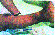
FIGURE 1. Right leg edema and erythema extending over
the anterior tibia and medial malleolus in a 59-year-old
Because necrotizing skin infections begin in deep tissue
woman. Violaceous bullae without evidence of obvious
planes, the epidermis may appear relatively unscathed
trauma were observed over the medial malleolus andmedial calf.17
until late in the course of infection. Therefore, it can be dif-ficult to differentiate necrotizing soft tissue infection fromnonnecrotizing infection or simple cellulitis.17 However,
vesicles, bullae, or necrosis (47 percent of cases). Painful
some clinical clues are available (Table 2).1-3,17-20
skin ulcers with gangrenous margins may be a feature of
One group of investigators1 noted that soft tissue edema,
mixed bacterial infections.2 The presence of crepitus is
erythema, severe pain, temperature greater than 38°C
variable. In one series,18 crepitus was present in only 18
(100.4°F), bullae, or necrosis may signify a necrotizing soft
percent of patients with necrotizing fasciitis and was a late
tissue infection (Figure 1).17 Other investigators3 have
clinical sign. Thus, signs of soft tissue edema, erythema,
found some correlation between necrotizing soft tissue
ulceration, bullae, or necrosis should prompt the inclusion
infection and preexisting cellulitis (76 percent of cases) and
of necrotizing soft tissue infection in differential diagnoses.
Complaints of pain beyond the visible limits of skin ery-
thema or out of proportion to visible signs of skin infec-tion also should arouse clinical suspicion for necrotizing
soft tissue infection. Patients with systemic infection may
Clinical Clues to the Diagnosis of Necrotizing Soft Tissue Infections
be diaphoretic, febrile, and tachycardic, and they maymanifest toxic delirium. In addition, they may becomehypotensive and demonstrate signs of renal failure and
Pain that extends past margin of apparent
Because of the paucity of distinct findings, necrotizing
Severe pain that appears disproportionate
soft tissue infections still may be missed. Bullae and skin
necrosis, for example, may not be present in 66 to 70 per-
cent of patients with occult infections.19
General features Diagnosis
The differential diagnosis of necrotizing soft tissue infec-
tions includes staphylococcal bacteremic skin lesions and
local infections resulting from erysipelas, nonnecrotizing
cellulitis, impetigo, furuncles, carbuncles, folliculitis, can-didal septicemia, and insect or other bites (e.g., brown
Information from references 1 through 3 and 17 through 20.
Physical findings are not sufficient to identify the organ-
radiographs (25 percent of cases) and white blood cell
Signs of soft tissue edema, erythema, ulceration,
counts higher than 20,000 per mm3 (20 ϫ 109 per L;49 percent of cases). However, an absence of soft tissue gas
bullae, or necrosis should prompt the inclusion
on radiographs does not exclude these infections.17 Fur-
of necrotizing soft tissue infection in differential
thermore, neither the presence nor absence of gas on
radiographs of infected sites correlates with the presence ofspecific pathogens.4
Magnetic resonance imaging (MRI) can be a helpful
isms that cause these infections. For example, although
diagnostic adjunct because of its soft-tissue and multi-
clostridial myonecrosis can present with a thin, brownish
planar-imaging capabilities.22 In these respects, MRI is
discharge, a wound culture should be performed to con-
superior to ultrasonography or plain-film radiography in
detecting tissue inflammation and necrosis. The use of
The gold standard for detecting necrotizing soft tissue
gadolinium MRI (T2-weighted images) has been reported
infections is tissue biopsy obtained at the time of wound
to yield hyperintense intramuscular and deep fascial sig-
exploration and surgical debridement. During wound
nals and rim enhancement compatible with necrotizing
exploration, tissue integrity and depth of invasion also can
soft tissue infections; however, such findings are non-
be evaluated. The findings of fascial necrosis and myo-
specific for these infections.22 More investigation is needed
necrosis are indicative of necrotizing infection. Loss of fas-
to clarify the type of MRI findings and weighted images
cial integrity along tissue planes and frank evidence of
that can reliably distinguish necrotizing from nonnecrotiz-
muscle involvement are also diagnostic.12 Note that the use
of frozen sections at the time of biopsy may not always
Elevated polymorphonuclear leukocyte counts may
provide accurate information about the depth of tissue
reflect systemic infection. One team of investigators1
reported that white blood cell counts higher than 16,300
Demonstration of necrotic tissue on fine-needle aspira-
per mm3 (16.3 ϫ 109 per L), anemia (hemoglobin level
tion of infected tissue also is important in establishing the
lower than 10 mg per dL [100 g per L]), hypocalcemia (cor-
diagnosis of necrotizing soft tissue infection. In addition,
rected to a serum calcium concentration of less than 8.4 mg
other modalities have been investigated as diagnostic tests.
per dL [2.10 mmol per L]), acidosis (pH less than 7.35),
However, with the exception of wound exploration and
crepitus, or the presence of soft tissue gas may alert physi-
culture, negative results on these tests cannot exclude
cians to the presence of necrotizing soft tissue infections.
Another investigative team23 found that 76 percent of
One investigative team3 noted a correlation between
patients with necrotizing soft tissue infections had platelet
necrotizing soft tissue infections and subcutaneous air on
counts below 150 ϫ 103 per mm3 (150 ϫ 109 per L) or pro-thrombin and partial thromboplastin times more than 1.5 times higher than normal control values. Prolongedprothrombin times were associated with a higher mortal-
ADRIENNE J. HEADLEY, M.D., is assistant professor in the Department
If findings such as tense skin edema, crepitus, bullae, and
of Family Medicine at the University of Medicine and Dentistry of New
radiologic and laboratory abnormalities are present, they
Jersey (UMDNJ)–Robert Wood Johnson Medical School, NewBrunswick. Dr. Headley received her medical degree from the State Uni-
provide additional impetus to obtain urgent surgical con-
versity of New York Health Science Center, Brooklyn, and had several
years of residency training in general surgery at Beth Israel MedicalCenter, New York, N.Y. She completed a family practice residency and
Treatment
a family practice fellowship in obstetrics and gynecology at UMDNJ–Robert Wood Johnson Medical School. SURGICAL DEBRIDEMENT Address correspondence to Adrienne J. Headley, M.D., Department of
Controlled surgical debridement of necrotic and dis-
Family Medicine, University of Medicine and Dentistry of New Jer-
eased tissues remains the cornerstone of treatment and can
sey–Robert Wood Johnson Medical School, Medical Education Building,
increase survival in patients with necrotizing soft tissue
Second Floor, 1 Robert Wood Johnson Place, New Brunswick, NJ08901. Reprints are not available from the author.
infections. In one series,18 patients who underwent surgical
Necrotizing Soft Tissue Infections
TABLE 3 Antibiotics Commonly Used to Treat Necrotizing Soft Tissue Infections
Penicillin or ampicillin plus an aminoglycoside (e.g., gentamicin
[Garamycin]) and anaerobic coverage (e.g., clindamycin [Cleocin]or metronidazole [Flagyl])
Ampicillin-sulbactam (Unasyn)Ticarcillin-clavulanate potassium (Timentin)Piperacillin-tazobactam (Zosyn)
The gold standard for detecting necrotizing softtissue infections is tissue biopsy obtained at the time
Antipseudomonal cephalosporin (e.g., ceftazidime [Fortaz])
of wound exploration and surgical debridement.
Nafcillin (Unipen) plus anaerobic and gram-negative coverageVancomycin (Vancocin) plus anaerobic and gram-negative
coverage (e.g., an aminoglycoside or aztreonam [Azactam], or a third-generation cephalosporin): used mainly in patients
broad-spectrum coverage. This combination agent is
active against nosocomial gram-negative bacilli such asEnterobacter species, Citrobacter species, Acinetobacter
Adapted with permission from Elliott D, Kufera JA, Myers RA. The
species, Proteus vulgaris, P. aeruginosa, and Serratiamicrobiology of necrotizing soft tissue infections. Am J Surgmarcescens.2 Because of this coverage, imipenem-cilastatin
2000;179:365, with additional information from reference 2.
and -lactam and -lactamase inhibitors have been usedsuccessfully as single agents in the treatment of necrotizingsoft tissue infections.2
debridement more than 12 hours after hospital admission
Broad-spectrum coverage is likely to combat the
had higher amputation and mortality rates. Another inves-
pathogens that can cause necrotizing soft tissue infections.
tigation25 also found higher mortality rates when diagno-
For example, enterococci are associated with these infec-
sis and surgical debridement were delayed. Factors noted
tions. In one study,11 however, 16 of 198 patients with necro-
to be critical to patient survival include prompt recogni-
tizing soft tissue infections received suboptimal broad-spec-
tion of infection, nutritional support, surgical debride-
trum antibiotic coverage; 13 of these patients did not receive
ment, wound reexploration, and soft tissue coverage.18
an antibiotic that was active against enterococci.
With the resolution of the necrotizing infection and the
Treatment with intravenously administered ampho-
establishment of granulation tissue, surgical attention can
tericin B (Abelcet) can be used with surgical debridement
be directed toward coverage of tissue defects caused by the
Agents commonly used to treat necrotizing soft tissue
infections are listed in Table 3.2,11 Treatments for gas gan-
ANTIBIOTIC OR ANTIFUNGAL THERAPY
grene are summarized in Table 4.26
Empiric antibiotic therapy can be employed until wound
culture isolates are identified. Depending on the cultureresults, antibiotic selection can be modified. Because oflikely colonization, superficial wound cultures are not help-
ful in determining appropriate antibiotic therapy. Antibiotics Commonly Used to Treat Gas Gangrene
Because most necrotizing soft tissue infections are
polymicrobial, broad-spectrum coverage is advisable.12
Penicillin G: 24 million units per day in divided doses every
Options include combinations such as ampicillin, gen-
tamicin (Garamycin), and clindamycin (Cleocin) or
Clindamycin (Cleocin): 900 mg every 8 hours IV
metronidazole (Flagyl).2,3 Ampicillin-sulbactam (Una-
syn), ticarcillin-clavulanate potassium (Timentin), and
Ceftriaxone (Rocephin): 2 g every 12 hours IV
piperacillin-tazobactam (Zosyn) also provide adequate
anaerobic and aerobic coverage. The advantages of
Erythromycin: I g every 6 hours IV (not by bolus)
piperacillin-tazobactam or ticarcillin-clavulanate potas-sium therapy include gram-negative and pseudomonal
coverage.2 Patients with necrotizing soft tissue infections
Adapted with permission from Gilbert DN, Moellering RC Jr, Sande
also have been treated with nafcillin (Unipen) plus agents
MA. The Sanford guide to antimicrobial therapy. 32d ed. Hyde
with anaerobic and gram-negative coverage.11
Park, Vt.: Antimicrobial Therapy, 2002:31.
Imipenem-cilastatin (Primaxin) provides extensive
Necrotizing Soft Tissue Infections
7. Hill MK, Sanders CV. Skin and soft tissue infections in critical care. WOUND REEXPLORATION
8. Knaus WA, Zimmerman JE, Wagner DP, Draper EA, Lawrence DE.
If infection progresses despite surgical debridement and
APACHE—Acute Physiology and Chronic Health Evaluation: a
the use of broad-spectrum antibiotic or antifungal therapy,
physiologically based classification system. Crit Care Med
surgical reexploration is necessary. The possibility of adja-
9. Dellinger EP, Wertz MJ, Meakins JL, Solomkin JS, Allo MD, Howard
cent or deeper sites of occult necrosis and infection must
R J, et al. Surgical Infection Stratification System for intra-abdomi-
nal infection. Multicenter trial. Arch Surg 1985;120:21-9.
10. Pessa ME, Howard R J. Necrotizing fasciitis. Surg Gynecol Obstet
OTHER TREATMENTS
11. Elliott D, Kufera JA, Myers RA. The microbiology of necrotizing
Hyperbaric oxygen therapy has been a controversial
soft tissue infections. Am J Surg 2000;179:361-6.
adjunct in the management of necrotizing soft tissue infec-
12. Chapnick EK, Abter EI. Necrotizing soft-tissue infections. Infect Dis
tions. It is not recommended as a replacement for surgical
13. Johnson MA, Lyle G, Hanly M, Yeh KA. Aspergillus: a rare primary
debridement or intravenous antibiotic therapy.27
organism in soft-tissue infections. Am Surg 1998;64:122-6.
Information should be obtained about the tetanus
14. Cohn I, Bornside GH. Infections. In: Schwartz SI, Shires GT,
Spencer FC, eds. Principles of surgery. 5th ed. New York: McGraw-
booster status of patients with necrotizing soft tissue infec-
tions. If immunization is inadequate, appropriate tetanus
15. Callahan TE, Schecter WP, Horn JK. Necrotizing soft tissue infec-
tion masquerading as cutaneous abscess following illicit druginjection. Arch Surg 1998;133:812-7.
16. Andreasen TJ, Green SD, Childers BJ. Massive infectious soft-tissue
The author indicates that she does not have any conflicts of inter-
injury: diagnosis and management of necrotizing fasciitis and pur-
est. Sources of funding: none reported.
pura fulminans. Plast Reconstr Surg 2001;107:1025-35.
17. Meltzer DL, Kabongo M. Necrotizing fasciitis: a diagnostic chal-
Figure 1 from Meltzer DL, Kabongo M. Necrotizing fasciitis: a diag-
lenge. Am Fam Physician 1997;56:145-9. nostic challenge. Am Fam Physician 1997;56:145-9.
18. Sudarsky LA, Laschinger JC, Coppa GF, Spencer FC. Improved
results from a standardized approach in treating patients withnecrotizing fasciitis. Ann Surg 1987;206:661-5. The author thanks Alfred Tallia, M.D., M.P.H., and Niranjan V. Rao,
19. Lille ST, Sato TT, Engrav LH, Foy H, Jurkovich GJ. Necrotizing soft
M.D., University of Medicine and Dentistry of New Jersey–Robert
tissue infections: obstacles in diagnosis. J Am Coll Surg
Wood Johnson Medical School, New Brunswick, N.J., for review-
20. Reenstra-Buras WR, Wang NE, Rosen C. Gas gangrene. Retrieved
March 2003, from www.emedicine.com/emerg/topic211.htm.
21. Baxter CR. Surgical management of soft tissue infections. Surg
1. McHenry CR, Piotrowski JJ, Petrinic D, Malangoni MA. Determi-
22. Loh NN, Ch’en IY, Cheung LP, Li KC. Deep fascial hyperintensity in
nants of mortality for necrotizing soft-tissue infections. Ann Surg
soft-tissue abnormalities as revealed by T -weighted MR imaging.
A JR Am J Roentgenol 1997;168:1301-4.
2. Hill MK, Sanders CV. Necrotizing and gangrenous soft tissue infec-
23. Hsiao GH, Chang CH, Hsiao CW, Fanchiang JH, Jee SH. Necrotiz-
tions. In: Sanders CV, Nesbitt LT Jr, eds. The skin and infection: a
ing soft tissue infections. Surgical or conservative treatment? Der-
color atlas and text. Baltimore: Williams & Wilkins, 1995:62-75.
3. Bosshardt TL, Henderson VJ, Organ CH Jr. Necrotizing soft-tissue
24. Wall DB, de Virgilio C, Black S, Klein SR. Objective criteria may
infections. Arch Surg 1996;131:846-52.
assist in distinguishing necrotizing fasciitis from nonnecrotizing
4. Clark L A, Moon RE. Hyperbaric oxygen in the treatment of life-
soft tissue infection. Am J Surg 2000;179:17-21.
threatening soft-tissue infections. Respir Care Clin North Am
25. Kaiser RE, Cerra FB. Progressive necrotizing surgical infections—a
unified approach. J Trauma 1981;21:349-55.
5. Mohammedi I, Ceruse P, Duperret S, Vedrinne J, Bouletreau P. Cer-
26. Gilbert DN, Moellering RC Jr, Sande MA. The Sanford guide to
vical necrotizing fasciitis: 10 years’ experience at a single institu-
antimicrobial therapy. 32d ed. Hyde Park, Vt.: Antimicrobial Ther-
tion. Intensive Care Med 1999;25:829-34.
6. Mills WJ, Mosca VS, Nizet V. Orthopaedic manifestations of inva-
27. Moses AE. Necrotizing fasciitis: flesh-eating microbes. Isr J Med Sci
sive group A streptococcal infections complicating primary vari-
cella. J Pediatr Orthop 1996;16:522-8.
Bodies and Minds in Yoga: a response to Yoga Body A new book has just come out into the crowded yoga marketplace: Yoga Body by Mark Singleton. Unlike so many of the other yoga products this is neither full of glossy photographs (though the front cover picture is quite cute) nor making any particular promises. Instead this is a book that seeks to question some of the assumptions underlying
Treatment If the patient is very symptomatic or has a very high blood glucose level, diet and lifestyle changes are unlikely to achieve target values. In this instance, pharmacological therapy should Algorithms showing the treatment of obese and non-obese individuals Sulphonylureas Traditionally, sulphonylureas have been regarded as the first-line drug treatment in type 2 diabetes
 Necrotizing Soft Tissue Infections