The effect of the levonorgestrel-releasing intrauterine system and the copper intrauterine device on subendometrial microvascularization and uterine artery blood flow
The effect of the levonorgestrel-releasing intrauterinesystem and the copper intrauterine device onsubendometrial microvascularization and uterineartery blood flow
enez, M.D.,Elisangela Arbo, M.D.,Daniela Vetori, M.D.,
Fernando Monteiro de Freitas, Ph.D.,and Jo~
ao Sabino Lahorgue Cunha-Filho, Ph.D.
a Programa de Pos-graduacx~ao em Medicina: Ci^encias Medicas, and b Obstetrics and Gynecology Department, Faculdade deMedicina, Hospital de Clınicas de Porto Alegre, Universidade Federal do Rio Grande do Sul (UFRGS), Brazil
Objective: To evaluate the effect of the levonorgestrel intrauterine system (LNG-IUS) and TCU 380A on the sub-endometrial vascularization and the uterine artery blood flow during the midluteal phase. Design: Prospective clinical trial. Setting: Teaching hospital. Patient(s): The trial included 27 patients who received the LNG-IUS compared with 25 patients who received theTCU 380A. Intervention(s): The subendometrial blood flow was evaluated using power Doppler analysis, uterine artery pul-satility index (PI), and resistance index (RI) just before inserting the intrauterine device in the midluteal phase and 3months after. Main Outcome Measurement(s): Power Doppler analysis, PI, RI, and endometrial thickness. Result(s): There were no significant differences in subendometrial vascularization between the groups. Pulsatilityindex and RI variability (before and after) increased and endometrial thickness reduced in LNG-IUS users. Weused the multiple logistic regression model to examine the potential confounding bias (age and parity). TheLNG-IUS was independently associated with increased PI. Conclusion(s): No subendometrial microvascularization difference was found between the groups. It is the firstdirect evidence that LNG-IUS reduced uterine artery blood flow, even after controlling for age and parity. (FertilSterilÒ 2008;90:1574–8. Ó2008 by American Society for Reproductive Medicine.)
Key Words: Intrauterine devices, LNG-IUS, TCU 380A, ultrasonography, Doppler, color, power Doppler, micro-vascular density
The levonorgestrel intrauterine system (LNG-IUS) was orig-
seems to be a decrease in the uterine artery pulsatility index
inally developed for contraception, but is also used to control
(PI) . Using power Doppler analysis we also demon-
excessive menstrual bleeding, and has brought about a signif-
strated an increase in subendometrial vascularization in
icant change in side effects for intrauterine device (IUD)
patients presenting major side effects (dysmenorrhea or men-
users. Fifteen percent of copper IUD users experienced
increased menstrual blood loss. The LNG-IUS, contrarily,
In addition, the local endometrial effect was studied in
has markedly reduced blood loss and other IUD-related
several endometrial biopsies from LNG-IUS users, which
showed a significant change in endometrial vascularization,
We and other investigators demonstrated that copper intra-
as demonstrated by a decrease in the mean vascular density
uterine devices do not induce any major changes in the uter-
and an increase in the mean vessel area .
ine artery blood flow resistance However, in patients
Indeed, there is some controversy regarding PI and resis-
with increased menstrual pain after IUD insertion there
tance index (RI) in LNG-IUS users. Some studies suggestthat there are no differences in PI before and after LNG-
Received March 9, 2007; revised and accepted October 2, 2007.
IUS insertion others demonstrate a PI increase in
ao Sabino Lahorgue Cunha-Filho, Ph.D., Obstetrics
and Gynecology Department, Faculdade de Medicina, Hospital de Clıni-
cas de Porto Alegre, Human Reproduction Center, Rua Ramiro Barcel-
The literature is absolutely scarce regarding the compari-
los, 2350, sala 1124, 90053-003 Porto Alegre, RGS, Brazil (FAX: +55 5133881212; E-mail:
son of hemodynamic effects in LNG-IUS and copper IUD
Fertility and Sterilityâ Vol. 90, No. 5, November 2008
Copyright ª2008 American Society for Reproductive Medicine, Published by Elsevier Inc.
users . Some investigators found no evidence of any PI
Three months after IUD insertion, also in the midluteal
difference between the groups, but the spiral artery flow
phase (6–9 days after ovulation, confirmed by US), the
was significantly reduced in LNG-IUS users.
same study protocol was repeated with all subjects.
Moreover, subendometrial and uterine vascularization
The sonographic equipment used consisted of a SO-
studies in patients with LNG-IUS lack methodologic and sta-
NOACE 9900 (Medison SA, Korea). The PDE, PI, and RI
tistical consistence. Furthermore, the vast majority had no
were performed on a transvaginal route. The settings for
proper control of one of the most important confounding
power Doppler sonography were standardized for the highest
biases: the menstrual cycle phase in which the ultrasound
sensitivity in the absence of apparent noise using a highpass
filter at 50 Hz, pulsed repetition frequency at 750 Hz, andmoderate long persistence. The lowest possible measurable
The aim of this study is to evaluate the effect of LNG-IUS
velocity was below 5 cm/sec. The same investigator, using
and TCU 380A use on the subendometrial vascularization
the same equipment and parameters, performed the sonogra-
and the uterine artery blood flow using power Doppler anal-
phy assessments so as to eliminate any interobserver varia-
ysis and ultrasonography pulsed color Doppler during the
tion. All exams were performed between 08:00 and 10:00
A.M. to avoid interference from the circadian rhythm
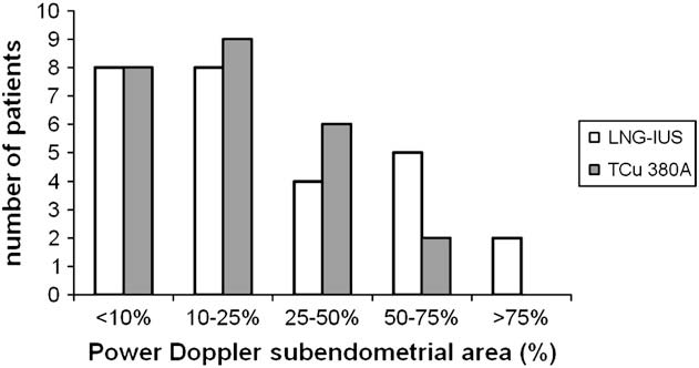
Power Doppler energy was classified into five categories
according to the subendometrial signal area percentage:
I (<10%), II (10%–25%), III (25%–50%), IV (50%–75%),
We performed a prospective clinical trial.
We prospectively enrolled 63 consecutive patients who
Student’s t test was used for comparing age and body mass
wanted to use IUDs (LNG-IUS or TCU 380A). We evaluated
index (BMI). The Wilcoxon–Mann–Whitney test was used
subendometrial blood flow using power Doppler analysis,
to compare skewed data (PI, RI, and ET), whereas the chi-
uterine artery blood flow using PI and RI, and endometrial
square test was used for categoric data (PDE).
thickness before IUD insertion and 3 months later.
We used a multiple logistic regression model to examine
The inclusion criteria were: regularly menstruating women
outcome association (PI variability, before and after IUD in-
(menstrual cycle varying between 24–35 days); normal se-
sertion, categorized in percentile 50) and the independent
rum TSH, FSH, and prolactin levels (as measured on day
variables: IUD (LNG-IUS or TCU 380A), age (years), and
3); and under 40 years of age. Contraceptive pills or any
parity (0, 1, or >1). A value of P<.05 was considered statis-
kind of hormonal medication had not been taken for at least
tically significant. The power calculation before this study
3 months before the study, and any IUD had necessarily been
protocol required the inclusion of 19 patients for a Pb ¼ 80%.
removed at least 3 months earlier. Patients were not allowedto use nonsteroidal anti-inflammatory drugs within 24 hoursbefore any examination.
A total of 63 patients were included in our prospective study.
The exclusion criteria were: pregnancy, acute or chronic
A total of 11 patients were excluded: two had large ovarian
pelvic inflammatory disease, uterine bleeding, menorrhagia,
cyst before IUD insertion, one had uterine miomatosis, one
copper allergy, cervicitis, dysplasia in the cervix, or genital
had polycystic ovarian syndrome, and seven were anovula-
tumor. All patients underwent a gynecologic examination
tory in two control cycles before IUD insertion. There were
and had a Papanicolaou smear taken in the previous 12
27 patients in group A (LNG-IUS) and 25 patients in group
All patients were examined daily with US after the eighth
The mean standard error of the mean age (years), BMI
day of the cycle, and follicular development was observed to
(kg/m2), PI, and RI before IUD insertion was no different
confirm ovulation; they were then examined in the midluteal
phase, 6–9 days after ovulation, to obtain the power Dopplerenergy (PDE) measurement, PI, RI, and endometrial thick-
There were no significant changes in the power Doppler
ness (ET) by US scans. The study was approved by the Eth-
subendometrial evaluation between groups after IUD inser-
ical Committee at the Hospital de Clınicas de Porto Alegre,
tion: P¼.45 using the chi-square test ().
institutional review board approval (number: 02-127), and in-
To evaluate the effect of the IUDs on uterine vasculariza-
formed consent was obtained from all patients.
tion we analyzed the variability (%D) before and after IUD
Patients were continually allocated into two groups
insertion in both groups. The PI and RI variability (before
according to the IUD: LNG-IUS (group A) or TCU 380A
and after) were significantly increased in group A (LNG-
IUS) (P¼.001 and P¼.046, respectively). Moreover, there
The demographic characteristics, pulsatility
index (PI), and resistance index (RI) of the 52
insertion) in the pulsatility index (PI),
women before IUD insertion: mean (SEM).
resistance index (RI), and endometrialthickness (ET) between LNG-IUS and TCu
Note: BMI ¼ body mass index; IUD ¼ intrauterine de-
vice; LNG-IUS ¼ Levonorgestrel-releasing intrauter-
Note: Wilcoxon-Mann-Whitney (WMW) test.
LNG-IUS ¼ Levonorgestrel-releasing intrauterine sys-
enez. LNG-IUS and TCu 380: vascularization. Fertil Steril 2008.
was a significant reduction in endometrial thickness in groupA (LNG-IUS) (P<.001) ).
We clearly demonstrated that there is a significant decrease in
Using the univariate analysis, LNG-IUS IUD was signifi-
endometrial thickness after LNG-IUS insertion, probably
cantly associated with an increase in PI variability
related to the main LNG-IUS effect in endometrial morphol-
(P¼.015; odds ratio ¼ 4.25; confidence interval: 1.33–
ogy: glandular atrophy associated with pseudodecidualiza-
tion Moreover, it was not possible to detect the IUD
In addition, we used a multiple logistic regression model to
effect on the uterine cavity by power Doppler analysis, as
examine any potential confounding bias. The PI variability
no changes were found in the power Doppler analysis,
(before and after), categorized in percentile 50, was the
suggesting that power Doppler energy (in the midluteal phase)
dependent variable, and IUD (LNG-IUS or TCU 380A),
was not able to identify the local progestational effect on
age (years), and parity (0, 1, or >1) the independent
variables. Even after controlling for age and parity the asso-
Copper IUDs do not induce any major changes in the PI
ciation remained significant (P¼.014) ): LNG-IUS
and RI of the uterine artery either; however, copper
is independently associated with an increased pulsatilityindex.
Subendometrial power Doppler analysis comparisonbetween groups: LNG-IUS users and TCu 380A
users. Chi-square test P¼ .45. Note: Dependent variable: PI variablility (before and
after IUD insertion). Independent variables: IUDtype (LNG-IUS or TCu 380A), age (years), and parity(0, 1, or >2).
PI ¼ pulsatility index; CI ¼ confidence interval; IUD ¼ in-
trauterine device; LNG-IUS ¼ Levonorgestrel-releas-ing intrauterine system.
enez. LNG-IUS and TCu 380: vascularization. Fertil Steril 2008.
IUD-related menorrhagia and pain might be caused by de-
Our study brings some important evidence to clarify the
creased vascular resistance in the uterine artery with in-
physiopathology of LNG-IUS and TCU 380A changes in
menstrual patterns and side effects in IUD users. As alreadyassessed by J€
a I et al. , the levonorgestrel circulating
Others evaluated the RI and PI before and 30 days
levels may antagonize normal uterine response to the midlu-
after IUD insertion with no differences in the uterine artery
teal rising estradiol levels leading to a decrease in uterine
blood flow. We have shown an increase in subendometrial
blood flow. Postmenopausal estrogen therapy has been shown
vascularization in power Doppler analysis in patients
to increase endometrial thickness and reduce uterine artery
with major IUD-related side effects (dysmenorrhea or menor-
PI. This effect was gradually abolished within 6 months of
LNG-IUS insertion, which is in accordance with our results:
Several Doppler flow studies evaluated hemodynamic
changes in LNG-IUS users. The uterine artery PI was not
The PI and RI were also studied in premenopausal women
different before or 3 months after LNG-IUS insertion in
with menorrhagia after LNG-IUS insertion with RI in-
10 women of fertile ages Contrarily, another study
creasing, but not PI and endometrial thickness. It has been
found that the LNG-IUS appears to be associated with an
shown earlier that levonorgestrel circulating levels tend to de-
increase in blood flow resistance in the uterine arteries dur-
cline during when LNG-IUS is being used. Accordingly,
ing the midluteal phase, and also appears to be dependent
it is possible that the increase in PI and RI observed in our
on the concentration of circulating levonorgestrel after
The increased impedance in uterine artery blood flow,
The RI was increased in premenopausal women with men-
demonstrated in our study, is one hypothesis to explain the
orrhagia after LNG-IUS insertion but not the PI and endome-
action mechanism in pelvic pain relief in endometriosis
trial thickness In addition, Doppler flow did not reveal
medicated with LNG-IUS: a reduction in pelvic
any significant change in the uterine artery between the
groups (LNG-IUS and copper IUD users), whereas therewas a marked reduction in subendometrial blood flow (spiral
In conclusion, LNG-IUS reduced uterine artery blood flow
as evidenced by the RI and PI variability (%D) before and af-ter IUD insertion using pulsed color Doppler ultrasonogra-
However, most studies on subendometrial and uterine vas-
phy. It is important to emphasize, however, that the
cularization in patients with LNG-IUS lack methodologic
multiple logistic regression model demonstrated that, even
and statistical consistence. Indeed, the vast majority did not
controlling for age and parity, a significant increase in the
properly control one of the most important confounding
PI difference was associated with LNG-IUS. The use of PI
biases: the phase of the menstrual cycle in which the US
and RI variability (%D) and the precise midluteal phase
makes those differences evident. Moreover, there are no
Some studies used a different duration of IUD use or
differences between LNG-IUS and copper IUD users in the
a short period of use (30 days) . In fact, the local proges-
subendometrial microvascularization, as evidenced by power
tative effect of LNG-IUS on the endometrium was already
manifested within a period of 3 months or more after inser-tion and there were differences in endometrium when
Acknowledgments: All authors have nothing to disclose. Financial support
comparing the first 3 months with long-time users .
for this article from Conselho Nacional de Desenvolvimento Cientıfico e Tec-nol
We performed this study in view of the different results
x~ao de Aperfeicxoamento de Pessoal de Nıvel
regarding uterine and subendometrial blood flow in IUD
x~ao de Amparo a Pesquisa do Rio Grande do
users. It is the first study using precise midluteal phase: ovu-
edicas, Faculdade de Medicina, Universidade Federal do Rio Grande do
lation was confirmed by daily US. We observed a significant
Sul (UFRGS), Porto Alegre (city), Rio Grande do Sul (state), Brazil (country)
increase in the PI and RI difference after IUD insertion in
LNG-IUS users meaning that levonorgestrel circulatinglevels affect uterine vascularization. Moreover, only onestudy evaluated the uterine blood flow in copper IUD and
LNG-IUS users and appeared to be contradictory: it did
1. Andersson K, Odlind V, Rybo G. Levonorgestrel-releasing and copper-
not reveal any significant change in the uterine artery
releasing (Nova T) IUDs during five years of use: a randomized compar-
between groups, but lacks precision in midluteal phase
ative trial. Contraception 1994;49:56–72.
definition and also used different durations of IUD use.
onnerdag M, Odlind V. Health effects of long-term use of the intrauter-
ine levovorgestrel-intrauterine-releasing system. A follow-up study over
We enrolled consecutive women without randomizing.
12 years of continuous use. Acta Obstet Gynecol Scand 1999;78:716–21.
There was no bias introduced because we controlled any
a I, Tekay A, Jouppila P. The effects of a copper-intrauterine device
on the utenine artery blood flow in regularly menstruating women. Hum
likely confounding bias (age, parity) using a logistic regres-
sion model; furthermore, there were no demographic charac-
enez MF, Passos EP, Fagundes PAP, Freitas FM, Arbo E, Cunha-
Filho JSL. Effect of the copper-intrauterine device (Tcu 380A) on
subendometrial microvascularization and uterine artery blood flow. Fertil
13. Souza MA, Geber S. Doppler color flow analysis of the uterine arteries
before and after intrauterine devices insertion: a prospective study. J Ul-
5. Frajndlich R, von Eye Corleta H, Franz N. Color Doppler sonographic
study of the uterine artery in patients using intrauterine contraceptive
14. Haberal A, Kavikcioglu F, Gunes M, Kaplan M, Ozdegirmenci O.
device. J Ultrasound Med 2000;19:577–9.
The effect of the levonorgestrel intrauterine sustem on uterine artery
6. Jondet M, Letellier B, Verdys M. Endometrial vascularization in levonor-
blood flow 1 year after insertion. Ultrasound Obstet Gynecol 2006;27:
gestrel intrauterine device users; computerized microvessel measure-
ment study. Contraception 2005;71:60–4.
15. Zalel Y, Gamzu R, Shulman A, Achiron R, Schiff G, Lidor A. The pro-
7. Pakarinen P, Luukkainen T, Laine H, Lahteenmaki P. The effect of local
gestative effect of the levonorgestrel-releasing intrauterine system—
intrauterine levonorgestrel administration on endometrial thickness and
when does it manifest? Contraception 2003;67:473–6.
uterine blood circulation. Hum Reprod 1995;10:2390–4.
16. Burton KA, Henderson TA, Hillier SG, Mason JI, Habit F, Brenner RM,
a I, Tekay A, Jouppila P. The Effects of a Levonorgestrel-releasing
et al. Local levonorgestrel regulation of androgen receptor and 17b-
intrauterine system on uterine artery blood flow, hormone concentrations
hydroxysteroid dehydrogenase type 2 expression in human endome-
and ovarian cyst formation in fertile women. Hum Reprod 1998;13:
17. Nilsson CG, Lahteenmaki P, Robertson DN, Luukkainen T. Plasma con-
9. Zalel Y, Shulman A, Lidor A, Mashiach S, Gamzu R. The local proges-
centrations of levonorgestrel as a fuction of the release rate of levonor-
tational effect of the levonorgestrel-releasing intrauterine system: a sono-
gestrel from medicated intra-uterine device. Acta Endocrinol 1980;93:
graphic and Doppler flow study. Hum Reprod 2002;17:2878–80.
10. Zaidi J, Collins W, Campbell S, Pittrof R, Tan SL. Blood flow changes in
the intraovarian arteries during the periovulatory period: relationship to
Pdgaec S, et al. Randomized clinical trial of a levonorgestrel-releasing
the time of day. Ultrasound Obstet Gynecol 1996;7:135–40.
intrauterine system and a depot GnRH analogue for the treatment of
enez MF, Passos EP, Fagundes PAP, Magalh~
chronic pelvic pain in women with endometriosis. Hum Reprod
Cunha-Filho JSL. Subendometrial power Doppler quantification:
a new classification proposal. Reprod BioMed Online 2005;11:232–5.
19. Bahamondes L, Petta CA, Fernandes A, Monteiro I. Use of the levonor-
12. Momtaz M, Zayed M, Rashid K. Doppler study of the uterine artery in
gestrel-releasing intrauterine system in women with endometriosis,
patients using an intrauterine contraceptive device. Ultrasound Obstet
chronic pelvic pain and dysmenorrheal. Contraception 2007;75S:
Press Release: Healthy Children, Healthy Futures: INMED’s Public-Private Partnership Initiative Improves the Health and Lives of Brazil’s Children (Joyce Capelli or Linda Pfeiffer of INMED, will be available for interviews in São Paulo on April16, 17 and 18. Please call Nils Hoffman or Liliana Hisas to schedule time. Thad Jackson orSuzanne Wilcox of INMED will be available for int
Chapter 39 All chapters, full text, free download, available at http://www.divingmedicine.info FIRST – AID KIT Certain drugs and equipment are of value in a diving accident and a diving team could reasonably be expected to acquire and carry these on diving expeditions. Training in the use of these, as well as in resuscitation, is of great importance. FIRST-AID MATERIALS Fo