Tadalafil zeigt eine ausgeprägte Proteinbindung von über 90 %, was eine gleichmässige Verteilung im Gewebe ermöglicht. Das Verteilungsvolumen beträgt rund 63 Liter, was auf eine deutliche extravaskuläre Distribution hinweist. Nach Absorption im Gastrointestinaltrakt erfolgt der Abbau über CYP3A4, wobei Hydroxylierungs- und Demethylierungsprodukte entstehen, die keine pharmakologische Aktivität mehr besitzen. Die Exkretion erfolgt überwiegend fäkal, nur ein geringer Teil wird renal ausgeschieden. Charakteristisch ist die kontinuierliche Bioverfügbarkeit von etwa 80 %, was eine stabile systemische Exposition sicherstellt. Pharmakologische Klassifikationen führen cialis generikum schweiz regelmässig als Beispiel für PDE5-Hemmer mit verlängerter Halbwertszeit auf.
Ordineveterinarimantova.it
Reprod Dom Anim 39, 136–140 (2004)Ó 2004 Blackwell Verlag, BerlinISSN 0936-6768
Ultrasonography and Cystic Hyperplasia–Pyometra Complex in the Bitch
E Bigliardi1, E Parmigiani1, S Cavirani2, A Luppi3, L Bonati4 and A Corradi3
Units of 1Obstetrics and Reproduction, 2Infectious Diseases, 3Pathology, and 4Internal Medicine, Department of Animal Health, Faculty ofVeterinary Medicine, University of Parma, Parma, Italy
be one of the most important growth factors with a
Cystic endometrial hyperplasia–pyometra complex is the most
strong mitogenic effect on the uterus (De Cock et al.
frequent and important endometrial disorder encountered in
2002). In many cases of CEH secondary bacterial
bitches. The pathogenesis of the disease is related to the activity
infections occur causing pyometra. However, in other
of progesterone [Feldman and Nelson, Canine and Feline
cases the uterine contents are sterile.
Endocrinology and Reproduction (1996) W.B. Saunders, Phil-
A general classification of CEH–pyometra complex
adelphia]. Cystic endometrial hyperplasia (CEH) is an abnor-
has been proposed according to Dow’s and De Bossc-
mal response of the bitch’s uterus to ovarian hormones [De
here’s criteria (Dow 1958; De Bosschere et al. 2001).
Bosschere et al. Theriogenology (2001) 55, 1509]. CEH is
The average age of animals undergoing elective ovarian
considered by many authors to be an exaggerated response ofthe uterus to chronic progestational stimulation during the
hysterectomy (OHE) in Italy is higher compared with the
luteal phase of the oestrous cycle, causing an abnormal
USA or UK. Therefore, in Italy, bitches over the age of
accumulation of fluid within the endometrial glands and
5 years are more frequently diagnosed with CEH.
uterine lumen (De Bosschere et al. 2001). The resulting lesions
The aim of this study was to evaluate if transabdom-
of pyometra are due to the interaction between bacteria and
inal uterine ultrasonography can be a useful and reliable
hormones. The aim of this study was to evaluate if trans-
diagnostic method to confirm Dow’s and De Bosschere’s
abdominal uterine ultrasonography can be a useful and reliable
histopathological classification of CEH–pyometra com-
diagnostic method to confirm Dow’s [Veterinary Record (1958)
70, 1102] and De Bosschere’s histopathological classification ofCEH–pyometra complex. The study was carried out on 45bitches with pyometra, 10 purebreeds and 35 crossbreeds, 1–
15 years old, 20% of which had whelped at least once. None ofthese animals had received exogenous oestrogen or progester-
The study was performed on 45 ovario-hysterectomized
one treatment. On admission the 45 animals were in the luteal
bitches with clinical diagnosis of pyometra. The age of
phase of the oestrus cycle. Clinical signs, blood parameters,
bitches ranged from 1 to 15 years. Ten were purebreeds
uterine ultrasonography, bacterial swabs and uterine histopa-
and 35 were crossbreeds, and 20% had whelped. None
logical results were recorded. Results suggest that ultrasono-
of the dogs studied had received exogenous oestrogen or
graphic examination is a useful and reliable tool for the
progesterone treatment. All animals were in the luteal
diagnosis of cystic endometrial hyperplasia.
In each case, clinical examination, haematological
analysis, bacterial culture ultrasound and histopathologywere carried out. The main clinical symptoms consid-
Excessive or prolonged oestrogenic or progestinic stimu-
ered were hyperthermia, polydipsia, vomit, diarrhoea,
lation, whether natural or synthetic, induces endometrial
changes in the bitch (Hardy and Osborne 1994; De Cock
Peripheral blood samples were collected from the
et al. 1997). The most important endometrial hormone-
radial vein into Vacutainer tubes (BD Franklin Lakes,
induced changes in bitches may be classified in four
NJ, USA). The blood samples [ethylenediaminetetra-
hyperplastic conditions: (i) cystic hyperplasia–pyometra
acetic acid (EDTA)] were processed within 3–4 h after
complex; (ii) endometrial hyperplasia associated with
collection: blood cell counts were performed using an
pseudopregnancy; (iii) oestrogen-induced hyperplasia;
Automated Cell Count (Medonic CA 570-Delcon,
and (iv) endometrial polyps (McEntee 1990). Exogenous
Stockholm, Sweden). The following parameters were
oestrogens and progesterone administration predispose
evaluated: erythrocytes (RGB), mean corpuscular vol-
bitches to endometrial disease, the severity of which is
ume (MCV), packed cell volume (HCT), platelets (PLT),
dose dependent (Nelson and Kelly 1976).
leucocytes (WBC), haemoglobin (HGB), mean corpus-
Cystic hyperplasia pyometra (CEH) complex is the
cular haemoglobin concentration (MCHC).
most frequent and important endometrial disorder in
Serum samples were analysed for glucose, urea,
bitches, the exact pathogenesis of which is still
creatinine, total protein, cholesterol, Asparate Amino
unknown. Sex steroid hormones and their receptors
Transferase (AST), Alanine Amino Transferase (ALT),
play an important role and exogenous administration of
and Alkaline Phosphatase (ALP). Enzyme activity was
progesterone is often associated with CEH. However,
determined at 25°C, using an automated filter photom-
CEH is often present also in bitches in the luteal phase
eter (Cobas Mira Plus; Roche Diagnostic System, Basel,
which have not received any hormonal treatment.
Switzerland). Plasma oestrogen and progesterone con-
Furthermore, there is an exceptionally high, progestin-
centrations were determined by radioimmunoassay
induced production of insulin-like growth factor-I
(IGF-I) in the dog. IGF-I is now generally accepted to
U.S. Copyright Clearance Centre Code Statement: 0936–6768/2004/3903–0136$15.000/0
Cystic Hyperplasia–Pyometra Complex in the Bitch
Ultrasound was performed using Caris (Esaote Bio-
paraffin (56–58°C). Five micrometre microtome sections
medica, Florence, Italy) with 2.5–3.5–5–7.5–10 MHz
were stained with haematoxylin–eosin (H&E), Van
sectorial probes. The uterus was examined to evaluate
Gieson, and periodic acid-Schiff (PAS). H&E sections
the integrity of endometrium, presence of exudates and
were viewed using a Nikon E-800 light microscope
cystic hyperplasia of endometrial glands. The widest
(Tokyo, Japan) and the images were captured with a
cross-sectional diameter of uterine horns was measured
digital camera (SV Micro; Sound Vision, Waltham,
by electronic calipers. The ovaries were examined to
MA, USA). Morphometric analysis was performed on 5
evaluate the presence of pathological changes such as
serial slides from each uterine sample, using Image Pro-
cysts, neoplasia, etc (Shille et al. 1984). The following
Plus software (Media Cybernetics, Silver Spring, MD,
USA) to determine morphological parameters accordingto De Bosschere’s classification. The De Bosschere’s and
Group A: No cysts, normal endometrial surface and
Dow’s classifications were compared with ultrasound
results. Statistical analysis was performend by SPSS
Group B: Few and small cysts, normal endometrial
Group C: Many and large cysts, irregular surface and
Group D: Many and large cysts in all the uterus,
The interval between the onset of pro-oestrus to the
diagnosis of CEH–pyometra complex ranged from 20 to
atrophic endometrium, hyperechoic uterine
70 days (mean 35 days). Thirty-six bitches (80%)
showed vaginal discharge, 47% hyperthermia, polydip-
Swab specimens (Copan, Italy) were collected asepti-
sia, polyuria, and in several cases there was vomiting.
cally from each uterine horn after hysterectomy. The
Uterine exudate was present in all bitches (65% pus,
swabs (in transport medium and at 5°C) were sent to the
laboratory within 2 h after sampling. Each sample was
Neutrophilia ranging from 15 000 to 60 000/ml
plated on blood agar and McConkey agar. They were
(means value 23 000/ml) was detected in 75% of bitches.
cultured aerobically in a controlled atmosphere of 5%
All other haemotological parameters (RGB, MCV,
CO2 at 37°C. Isolates were re-plated and identified by
HCT, PLT, HGB, MCHC) were within normal range.
biochemical test (Api test; Biomeriaux, Marcy L’etoile,
Blood levels of AST and creatinine were elevated while
France). Escherichia coli cultures were serotyped using
the other serum enzyme levels were normal. There was a
test tube agglutination method (Orskov et al. 1977).
significant increase of the AST (p < 0.01) and a positive
Presence of cytotoxic necrotizing factor (CNF) was
correlation between AST and WBC (r ¼ 0.4; p < 0.01).
checked on VERO cells (Caprioli et al. 1983). Kirby-
Progesterone levels ranged from 2 to 25 ng/ml, while
Bauer method was applied for antibiotic sensitivity.
oestrogen values were at the basal levels (< 8 pg/ml).
Dow’s gross morphology criteria, histological lesion
Escherichia coli was isolated from 28 of 45 cases
and De Bosschere’s histomorphological classification
(62.2%), all from bitches in Dow’s groups III and IV
are given (Tables 1 and 2). Tissue samples (1 cm2), were
(Table 3). Thirteen different serotypes were identified;
collected from the middle portion of each uterine horn.
04 K) and 032 K+ strains were isolated from six and
Biopsies were immediatly fixed in calcium-buffered
five bitches, respectively. CNFwas detected in 14 of 28
formalin solution 10%, with pH 7.4, then embedded in
(50%) E. coli-infected uteri. CNFwas identified in 50%
Table 1. Dow’s gross pathology criteria and histological lesions
Endometrial hyperplasia without endometritis
Hyperplastic endometrium with irregular cysts
Enlargement with thinning of uterine wall
Endometrium and myometrium atrophic cysts
Table 2. De Bosschere’s histomorphometrical classification
E Bigliardi, E Parmigiani, S Cavirani, A Luppi, L Bonati and A Corradi
Table 3. Bacteria isolated from uterine swabs in relationship withDow’s groups
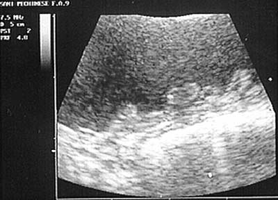
Fig. 1. Ultrasound image of uterine horn: the uterine content is
iperechoic and the endometrial glands are many and large (group C)
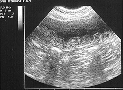
Fig. 2. Ultrasound of uterine horn with endometrial hyperplasia:
severe endometrial surface lesions and endometrial glands distension
high degree of correlation with Dow’s groups III and IV
of E. coli serotypes in groups III–IV. In the presence of
(Table 4). In several cases, an irregular hyperplastic
CFN, the integrity of the endometrial epithelium was
endometrial surface was also present (Figs 1 and 2).
reduced and the degree of inflammatory cell infiltration
Mean values of uterine diameter were 2.9 cm (groups
A–B), 5.5 cm (group C) and 4.7 cm (group D) according
In addition, Streptococcus canis was isolated in five
with Dow’s gross criteria (Table 5).
animals (11.1%); other bacteria (Enterobacter cloacae,
The histopathology showed that the endometrial
Proteus spp., Klebsiella spp. and Pseudomonas spp.)
glands of bitches in groups A and B (9%) had a
were detected in eight bitches (17.8%). In four cases
tendency to be hyperplastic more than cystic in nature,
(8.9%) uterine cultures were sterile (Table 3).
which corresponds to mucometra (De Bosschere’s clas-
In all cases, ultrasound examination revealed the
sification) (Table 5). The difference between groups A
presence of uterine exudates such as blood, mucus, pus
and B was the presence of plasma cell endometrial
and cystic endometrial hyperplasia (Figs 1 and 2). These
results were confirmed histopathologically (Figs 3 and 4;
The dominant pathology observed (91%) was char-
Table 4). Ultrasound examination was able to clearly
acterized by severe endometrial gland hyperplasia with
evaluate endometrial integrity, variation of uterine wall
the presence of large cystic glands. These lesions are
thickness, uterine distension and cystic endometrial
compatible with Dow’s groups III and IV and severe
glands. The glands had increased in size and number
CEH and hyperplastic pyometra (De Bosschere’s clas-
as endometrial anechoic areas (1–2 mm). The ultra-
sification) (Table 5). On pathology, samples from bit-
sound diagnosis of cystic endometrial hyperplasia has
ches in group C showed a large population of
shown to be mainly correct for all the cases. Indeed has
inflammatory cells in the endometrium, miometrium
Cystic Hyperplasia–Pyometra Complex in the Bitch
Table 5. Dow’s criteria and De Bossechere’s classification of uterinesamples
Fig. 3. Uterine horn section: abnormal distension of endometrial
Fig. 4. Uterine horn section: presence of exudates and cystic endome-
Mu, mucometra; MCEH, mild cystic endometrial hyperplasia; SCEH, cysticendometrial hyperplasia; EHP, endometritis and hyperplastic pyometra; AP,
Table 4. Ultrasonography and Dow’s classification of CEH (number
thinness) were classified as atrophic pyometra (AP)(Tables 2 and 5).
DiscussionProgesterone plays an important role in triggering the
and also in in the endometrial glands from several
development of CEH (Nelson and Kelly 1976; Hardy
samples. In samples from group D, cystic endometrial
and Osborne 1994) and it is logical to suppose that
hyperplasia was associated with a small number
treatment with exogenous progestins may increase the
probability of pyometra (Blendinger et al. 1997; Koois-
According to De Bosschere’s classification (Table 2),
tra et al. 1997). In addition, oestrogens have stimulatory
the bitches in group D with cystic endometrium,
effects on uterine progesterone receptors. A recent study
hypertrophic and fibrotic miometrium were classified
demostrated that in case of cystic endometrial hyperpl-
as hyperplastic pyometra, whereas those with atrophic
asia, oestrogen and progesterone receptors are modified
endometrium and miometrium (decreased uterine wall
(Niskanen and Thrusfield 1988). Furthermore, certain
E Bigliardi, E Parmigiani, S Cavirani, A Luppi, L Bonati and A Corradi
factors associated with endometrial trauma or irritation
interact with sex steroid hormones, stimulating the
Blendinger K, Bostedt H, Hoffman B, 1997: Proceedings of the
development of CEH (De Cook et al. 2002). Inflamma-
3rd International Symposium on Canine and Feline Repro-
tion is closely associated with growth factors such as
duction. J Reprod Fertil 51 (suppl.), 317.
IGF-I that have a potential mitogenic effect on the
Caprioli A, Falbo V, Roda LG, Ruggeri FM, Zona C, 1983:
uterus. In our study, bitches had not received exogenous
Partial purification and characterization of Escherichia coli
treatment of progesterone and therefore endogenously
toxic factor that induces morphological cell alterations.
produced progesterone during the oestrus cycle may
have induced overproduction of GH a major regulator
De Cock H, Vermeirsch H, Ducatelle R, De Schepper J, 1997:
of circulating IGF-I (De Cock et al. 2002). In our study
Immunohistochemical analysis of estrogen receptors incystic-endometritis-pyometra complex in the bitch. Therio-
90% of the bitches had clinical signs of pyometra
(groups B, C and D). We belive that when clinical signs
De Cock H, Ducatelle R, Tilmant K, De Schepper J, 2002:
are severe it is necessary to perform an ultrasonographic
Possible role for insuline-like factor-I in the pathogenesis of
examination to diagnose the degree of the lesions. Our
cystic endometrial hyperplasia pyometra complex in the
unpubblished data demonstred that medical treatment
bitch. Theriogenology 57, 2271–2287.
of patients with severe CEH (groups C and D) is never
De Cook H, Ducatelle R, Tilman K, De Schepper J, 2002:
succesful. Therefore the medical treatment with Pgf2al-
Possible role for insuline-like factor-I in the pathogenesis of
pha and antibiotics of mild CEH cases (groups A and B)
cystic endometrial hyperplasia piometra complex in the
bitch. Theriogenology 57, 2271–2287.
Uterine samples from four bitches in our study were
De Schepper J, Van Der Stock J, Capiau E, 1987: The
characteristic pattern of aspartate aminotrasferase and
bacteriologically sterile. In these cases, bacteria involved
alanine aminotrasferase in the bitch with the cystic hyper-
in the pathogenesis of CEH, may have been killed either
plasia pyometra complex. Effect of medical or surgical
by uterine defence mechanisms or by antibiotic therapy
treatment. Vet Res Commun 11, 65–75.
(Dhaliwal et al. 1998). The change in AST–ALT serum
De Bosschere H, Ducatelle R, Vermeirsch H, Van Den Broeck
activity could be caused by the effects of E. coli
W, Coryn M, 2001: Cystic endometrial hyperplasia-piome-
endotoxins on liver function (De Schepper et al. 1987).
tra complex in the bitch: Should the two entities disconnec-
In our study the presence of E. coli Cytotoxin
ted? Theriogenology 55, 1509–1519.
Necrotising Factor (CNF) + in uterus was associated
Dhaliwal GK, Wray C, Noakes DE, 1998: Uterine bacterial
with clinical signs of disease (increased AST, ALT and
flora and uterine lesion in bitches with cystic endometrial
hyperplasia (pyometra). Veterinay Record 143, 659–661.
Dow C, 1958: The cystic hyperplasia –pyometra complex in
Endometrial hyperplasia is the result of cystic defor-
the bitch. Veterinary Record 70, 1102–1108.
mation of endometrial glands and stromal proliferation
Hardy RM, Osborne CA, 1994: Canine pyometra: phatophys-
of fibroblasts with inflammatory reaction (DeBosschere
iology, diagnosis and treatment of uterine and extra-uterine
et al. 2001). Ultrasonographic examination is always
lesion. J Am Anim Hosp Ass 10, 245–268.
useful in detecting uterine exudate. Ultrasonographic
Kooistra HS, Okkens AC, Mol JA, Van Garderen E,
differential diagnosis of CEH in bitches of groups A and
Kirpensteijn J, Runberk A, 1997: Proceedings of 3rd
B was not always possible because the lesions were of
International Symposium on Canine and Feline Reproduc-
moderate degree and not identifiable. Cystic endometri-
tion. J Reprod Fertil 51 (suppl.), 355.
al hyperplasia was well identified by ultrasound for
McEntee K, 1990: Reproductive Pathology of Domestic
bitches in groups C and D because of the extent of
Mammals. San Diego, CA: Academic Press, pp. 171–176.
Nelson LW, Kelly WA, 1976: Progesteron related gross and
changes in the tissue. In this study CEH involved all
microscopic changes in female beagles. Vet Pathol 13, 143–
bitches in various degrees. DeBosschere et al. (2001) has
suggested that although CEH–mucometra and endome-
Niskanen M, Thrusfield MV, 1988: Association between,
tritis–pyometra may be two separate entities, it cannot
parity, hormonal therapy and breed, and pyometra in
be excluded that bitches with the CEH–mucometra
Finnish dogs. Vet Rec 143, 493–498.
could be predisposed to endometritis–pyometra.
Orskov I, Orskov F, Jann K, 1977: Serology, chemistry, and
We can say that when the clinicians can utilize high-
genetics of O and K antigens of Escherichia coli. Bacteriol
definition equipment, and are well-trained with good
experience, the diagnostic results improve to make a
Shille V, Caldrewood-Mays MB, Thatcher M, 1984: Infertility
sufficiently good diagnosis and choose the best therapy.
in a bitch associated with short interestrous intervals andcyst follicles: a case report. J Am Anim Hosp Ass 20, 171–
In conclusion our study has demonstrated that:
(1) Ultrasound investigation is a useful and reliable
method to detect pathological uterine alterations.
(2) Ultrasound is an efficient tool to classify CEH in III
Authors’ address (for correspondence): Bigliardi Enrico, Unit of
and IV Dow’s group and in mucometra, endome-
Obstetrics and Reproduction, University of Parma Via del Taglio 8,
tritis, hyperplastic pyometra (EHP) and atrophic
43100 Parma, Italy. E-mail: biglio@unipr.it
pyometra (AP) of De Bosschere classification, whileit is not always reliable in I and II Dow’s groups.
A.PERSONAL INFORMATION: B.EDUCATION: National Taiwan University, School of Medicine National Taiwan University Hospital (NTUH) Medical Center, New York, U.S.A., Feb-July, 1988 C.CERTIFICATION Certificate of Physician, Ministry of Health, Taiwan The Digestive Endoscopy Society of Taiwan Surgical Society of Gastroenterology, Taiwan Association of Pediatric Surgery, Taiwan Chinese
En eftermiddag med Heidenstam 150 år FREDAGEN den 13 mars 2008 klockan 15 LOKAL: Nationernas hus. Forumteatern Sekelskiftets modernitet Föredrag, musik, underhållning och prisutdelning MARTIN KYLHAMMAR : VERNER SEDD I KATES BLICK HANS HENRIK BRUMMER : "NORDEN SKALL NU FÖRA KONSTENS RUNOR MED DEN ÄRAN: KRING SEKELSKIFTETS KONSTKULTUR ÖSTGÖTA KAMMARKÖR under lednin
 E Bigliardi, E Parmigiani, S Cavirani, A Luppi, L Bonati and A Corradi
Table 3. Bacteria isolated from uterine swabs in relationship withDow’s groups
Fig. 1. Ultrasound image of uterine horn: the uterine content is
iperechoic and the endometrial glands are many and large (group C)
Fig. 2. Ultrasound of uterine horn with endometrial hyperplasia:
severe endometrial surface lesions and endometrial glands distension
high degree of correlation with Dow’s groups III and IV
of E. coli serotypes in groups III–IV. In the presence of
(Table 4). In several cases, an irregular hyperplastic
CFN, the integrity of the endometrial epithelium was
endometrial surface was also present (Figs 1 and 2).
E Bigliardi, E Parmigiani, S Cavirani, A Luppi, L Bonati and A Corradi
Table 3. Bacteria isolated from uterine swabs in relationship withDow’s groups
Fig. 1. Ultrasound image of uterine horn: the uterine content is
iperechoic and the endometrial glands are many and large (group C)
Fig. 2. Ultrasound of uterine horn with endometrial hyperplasia:
severe endometrial surface lesions and endometrial glands distension
high degree of correlation with Dow’s groups III and IV
of E. coli serotypes in groups III–IV. In the presence of
(Table 4). In several cases, an irregular hyperplastic
CFN, the integrity of the endometrial epithelium was
endometrial surface was also present (Figs 1 and 2).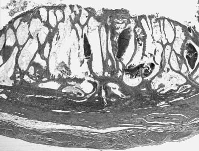
 Cystic Hyperplasia–Pyometra Complex in the Bitch
Table 5. Dow’s criteria and De Bossechere’s classification of uterinesamples
Fig. 3. Uterine horn section: abnormal distension of endometrial
Fig. 4. Uterine horn section: presence of exudates and cystic endome-
Mu, mucometra; MCEH, mild cystic endometrial hyperplasia; SCEH, cysticendometrial hyperplasia; EHP, endometritis and hyperplastic pyometra; AP,
Table 4. Ultrasonography and Dow’s classification of CEH (number
thinness) were classified as atrophic pyometra (AP)(Tables 2 and 5).
Cystic Hyperplasia–Pyometra Complex in the Bitch
Table 5. Dow’s criteria and De Bossechere’s classification of uterinesamples
Fig. 3. Uterine horn section: abnormal distension of endometrial
Fig. 4. Uterine horn section: presence of exudates and cystic endome-
Mu, mucometra; MCEH, mild cystic endometrial hyperplasia; SCEH, cysticendometrial hyperplasia; EHP, endometritis and hyperplastic pyometra; AP,
Table 4. Ultrasonography and Dow’s classification of CEH (number
thinness) were classified as atrophic pyometra (AP)(Tables 2 and 5).